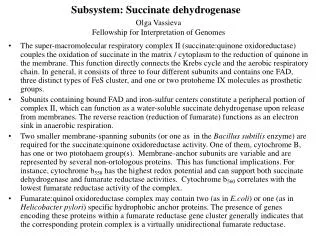
Subsystem: Succinate dehydrogenase
Olga Vassieva Fellowship for Interpretation of Genomes . Subsystem: Succinate dehydrogenase.

Subsystem: Succinate dehydrogenase
E N D
Presentation Transcript
Olga Vassieva Fellowship for Interpretation of Genomes Subsystem: Succinate dehydrogenase • The super-macromolecular respiratory complex II (succinate:quinone oxidoreductase) couples the oxidation of succinate in the matrix / cytoplasm to the reduction of quinone in the membrane. This function directly connects the Krebs cycle and the aerobic respiratory chain. In general, it consists of three to four different subunits and contains one FAD, three distinct types of FeS cluster, and one or two protoheme IX molecules as prosthetic groups. • Subunits containing bound FAD and iron-sulfur centers constitute a peripheral portion of complex II, which can function as a water-soluble succinate dehydrogenase upon release from membranes. The reverse reaction (reduction of fumarate) functions as an electron sink in anaerobic respiration. • Two smaller membrane-spanning subunits (or one as in the Bacillus subtilis enzyme) are required for the succinate:quinone oxidoreductase activity. One of them, cytochrome B, has one or two ptotohaem group(s). Membrane-anchor subunits are variable and are represented by several non-ortologous proteins. This has functional implications. For instance, cytochrome b558 has the highest redox potential and can support both succinate dehydrogenase and fumarate reductase activities. Cytochrome b560 correlates with the lowest fumarate reductase activity of the complex. • Fumarate:quinol oxidoreductase complex may contain two (as in E.coli) or one (as in Helicobacter pylori) specific hydrophobic anchor proteins. The presence of genes encoding these proteins within a fumarate reductase gene cluster generally indicates that the corresponding protein complex is a virtually unidirectional fumarate reductase.
Subsystem: Succinate dehydrogenase (SDH) SDH membrane anchor proteins SDH cytochrome B subunits
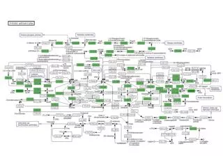
Examples of subsystem functional variantsin different genomes ? ? Variant codes: 1-4 subunit succinate dehydrogenase (2 catalitic+2 anchor subunits) 2-4 subunit fumarate reductase 3-4 subunit succinate dehydrogenase and 4 subunit fumarate reductase 4-3 subunit succinate dehydrogenase and 3 subunit fumarate reductase 5-3 subunit succinate dehydrogenase (or, in some cases, fumarate reductase?) 6-4 subunit succinate dehydrogenase and 3 subunit fumarate reductase 7-3 subunit fumarate reductase Fumarate reductase: presence of specific anchor subunits in gene cluster distinguishes it Quinone:Succinate Oxidoreductase
Subsystem Spreadsheet (fragment) Specific functional roles were assigned to different membrane anchor subunits of the complex: ? ? New putative variant of an anchor protein from Nostoc Cytochrome B-556 Heterodisulfide Reductase homolog Cytochrome B-558 Cytochrome B-? Cytochrome B-560 the E. coli variant of anchor protein
Occurrence of various membrane anchor subunits in different organisms Color coding: Eucarya, Rickettsia, Xylella, Xanthomonas, etc Escherichia, Pseudomonas, Mycobacteria, etc Sulfolobus, Synechocystis, Nostoc, Chlorobium Corynebacteria,Halobacterium, Prochlocococci,Thermoplasma, etc Bacilli, Staphylococci, Chlamidia, etc Methanobacteria Archaea Conserved subunits
Open questions, comments, conjectures 1. Missing genes • Succinate dehydrogenase anchor protein is missing in some organisms. Several gene candidates for this role identified in this study (as hypothetical membrane proteins clustered with known succinate dehydrogenase genes) have been included in the subsystem as “Hypothetical succinate dehydrogenase membrane anchor proteins”. However, anchor protein is still missing in cyanobacteria (should it be there at all?) • There are no NCBI records available pertaining to cyanobacterial Succinate dehydrogenase cytochrome b subunit. We were able to predicted an alternative form based on long-range homology in Prochlorococci, Synechococcus, Chlorobium and some other bacteria. • Synechocystis, Nostoc, Crocosphaera, Trichodesmium and Thermosynechococcus posess the second putative candidate for the role of Succinate dehydrogenase cytochrome b subunit, homologous to a Sulfolobus protein formerly annotated as Heterodisulfide reductase -- see next slide.
Open questions, comments, conjectures 2. Succinate dehydrogenase and Heterodisulfide reductase evolutionary interplay 30%Homology toHeterodisulfide reductase (Methanobacteria) In Methanobacteria HS-CoB and HS-CoM are direct soluble electron donors for fumarate reductase HS-CoB and HS-CoM. CoB-S-S-CoM Fumarate Succinate
At some point during evolution the Heterodisulfide reductase has probably formed a complex with functionally relevant catalytic subunits of fumarate reductase. Disappearance of the heterodisulfide reduction pathway (as a result of the switch from methanogenesis?) lead to further evolution of this protein into a specialized succinate dehydrogenase subunit, as the one now present in Sulfolobus and cyanobacteria. Succinate dehydrogenase and Heterodisulfide reductase evolutionary interplay continued Fig1 from From Iwasaki et al, J. Biol. Chem., Vol. 277, 42, 39642-39648 A, modular subunit arrangements of selected bacterial Frd complexes, S. tokodaii SdhABCD complex, and subunits of related enzymes (thiol:fumarate oxidoreductase and heterodisulfide reductase) from methanogenic archaea. B, the common cofactor arrangement (FAD and three FeS clusters) in bacterial FrdAB subcomplexes based on the reported crystal structures (5, 6). The FrdA/SdhA subunit contains the dicarboxylate active site at a covalently linked FAD, and the FrdB/SdhB subunit contains a high potential [2Fe-2S] cluster (Center S-1), a low potential [4Fe-4S] cluster (Center S-2), and a high potential [3Fe-4S] cluster (Center S-3) (1, 2, 4). The [3Fe-4S] cluster is replaced by a lower potential [4Fe-4S] cluster in the S. tokodaii SdhB subunit (13).