
Introduction
E N D
Presentation Transcript
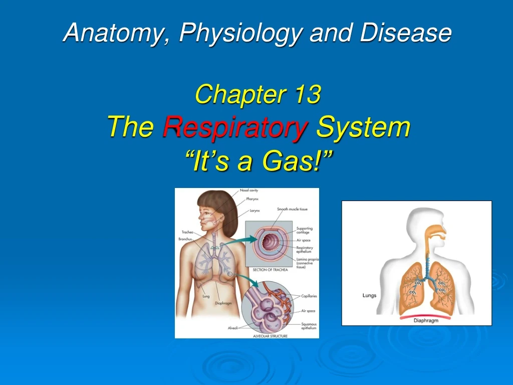
Health Science & OccupationsAnatomy, Physiology and Disease Chapter 13The Respiratory System“It’s a Gas!”
Introduction Respiratory systempurpose • to transport oxygen from environment and get it into blood stream to be utilized by cells. • moves 12,000 quarts of air per 24 hrs • removes waste gas – or carbon dioxide – from body to avoid “hyper-carbia.” • closely related to cardio-vascular system and they are sometimes grouped together as the cardio-pulmonary system.
Components include heart, blood, and network of blood vessels. Arteries carry blood away from heart, branch into smaller vessels called arterioles, which become capillaries, where nutrients are exchanged; capillaries become venules, that enlarge and become veins. System Overview
Components of Respiratory System • Two lungs that serve as vital organs • Upper and lower airways that conduct, or move, gas through system. • Terminal air sacs called alveoli surrounded by network of capillaries that allow gas exchange. • Thoracic cage that houses, protects, and facilitates function for system. • Muscles of breathing
“Air” contains many gases… • Nitrogen (78.08%) which is a support gas that keeps lungs open by adding volume, or filler, to vitally needed oxygen • Oxygen (20.95%)essential to life • Carbon Dioxide (0.03%) found in very small concentrations • Argon (0.93%) • Neon & Krypton: trace amounts
Ventilation vs. Respiration • Ventilation: is bulk movement of air down to terminal air sacs, or alveoli, of lungs. • Respiration: the process of gas exchange, where oxygen is added to blood and carbon dioxide is removed. • External Respiration: Movement of oxygen from alveoli to blood. • Internal Respiration: Movement of oxygen from blood to cells.
Boyle’s Law (PV=k1): when temp is constant so is pressure & volume. Charles’ Law (V=k2T): when pressure is constant so is volume. “Gas Law’s”
The Airways and Lungs • Human reserve oxygen: 4 to 6 minutes • Respiratory system is series of branching tubes called bronchi. • As branches get smaller they are called bronchioles & end in alveoli, terminal or distal end of respiratory system. • alveolus is surrounded by alveolar-capillary membrane & provides interface between respiratory and cardiovascular systems
Upper Airways • begin at nostrils, or nares, & end at vocal cords. • Functions: 1. heat/cool air 2. filtering & humidifying 3. olfactation (to smell) 4. phonations (produce sound) 5. ventilation: or conducting gas to lower airways.
Pathology Connection: • Allergic Rhinitis: DX: when allergens (like pollen) trigger nasal mucosa to secrete excessive mucous. S/S: runny nose, itchy, red or edematous eyes Rx: antihistamines
Pathology Connection: • Nasal polyps: DX: non-cancerous growths within nasal cavity S/S: chronic inflamation, dyspnea, nocturnal apnea Rx: surgically removed if they become large enough to block nasal passageway
Mucociliary Escalator • Nasal Cilia beat 1,000–1,500 times/min • propel gel layer & its trapped debris upward 1 inch/min to be expelled. • smokingparalyzes this escalator
Paranasal Sinuses • air-filled cavities found around nose • prolong and intensify sound • warm & humidify air • Not born with them: develop over time resulting in reformation of face and head.
Pharynx • hollow muscular structure starting behind nasal cavity, lined with epithelial tissue. • divided into 3 sections - nasopharynx - oropharynx - laryngopharynx
Nasopharynx • contains lymphatic tissue called adenoids; passageways into middle ear called Eustachian tubes.
Oropharynx • center section of pharynx • located behind oral, or buccal cavity • air, food and liquid, from oral cavity pass through • Contains tonsils • During swallowinguvula and soft palate move in posterior and superior position to protect nasal pharynx from entry of food or liquid
Laryngopharynx • Connects to both larynx, part of respiratory system, and esophagus, part of digestive system • Both food & air pass through • Potenial problem: - airway obstruction - infection - trauma
Larynx (voice box) • Semi-rigid structure composed of cartilage provide movement of vocal cords to control speech. • “Adams apple” (thyroid cartilage) is largest of cartilages found in larynx. • Cricoid cartilage lies below providing structure & support in exposed area of airway to prevent collapse. • Food travels into esophagus; air travels into larynx. • Glottis is opening that leads into larynx, & eventually lungs • Epiglottis: closes tightly when we swallow to prevent food from entering lungs
Pathology Connection • Common cold Etiology: over 200 different types of viruses Dx: acute inflammation of upper respiratory mucous membranes Rx: managing symptoms: antipyretics, antihistamines. - can be prevented with good hand-washing - not an allergy or influenza
Sinusitis Dx: Infection & inflammation of sinuses Etiology: chemical irritation vs bacterial S/S: pressure, pain, fever & headaches Rx: antipyretics, anti-inflammatory meds, antibiotics if bacterial not viral.
Tonsillitis Dx: Inflammation of tonsils Etiology: bacterial S/S: pain, dysphasia, fever, edema Rx: antibiotics, antipyretics, possible tonsillectomy.
Pharyngitis Dx: sore throat Etiology: Bacterial frequently Strep throat S/S: similar to Tonsillitis but with edema to neck glands. Rx: warm salt-H2O gargle antipyretics/anti- inflammatory meds, antibiotics if severe.
Laryngitis Dx: viral inflammation of voice box S/S: hoarseness Etiology: excessive use of voice Rx: complete voice rest, humidification
Acute Epiglotitis Dx: Dangerous infection causes swelling of epiglottis and airway obstruction. Etiology: 1. usually Haemophilus influenzae type B 2. most common in children 2-6 y/o 3. incidence lower when Flu shot taken S/S: fever, sore throat, respiratory distress, drooling, dysphasia, and dysphonia.
Acute Epiglottis, cont’d Rx: - onset is fast, & requires rapid treatment - maintain open airway - cool humidified O2 - orotracheal intubation or cricothyroidotomy - IV antibiotics, anti-inflammatory meds - hospitalization
Laryngotracheobronchitis“Croup” Dx: infection of laryngeal area Etiology: viral or bacterial S/S: barking cough like a goose, inspiratorystridor Rx: rest, antibiotics & anti-inflammatory meds Note: Sometimes called “Croup” or “Pertussis”
Trachea • Largest pipe in respiratory system • Begins bifurcating at center of chest into left and right mainstem bronchi @ carina. • Each bronchi branch into lobular bronchi that correspond to five lobes of lungs (3 in right; 2 in left) Lobes Upper Middle Lower Lobes Upper Lower
Epithelial Layers • First containsmucociliary escalator • Middle is lamina propria layer which contains smooth muscle, lymph, and nerve tracts • Third layer is protective and supportive basement cartilaginous layer Epithelial Layers First Middle Third
Bronchi • Branching continues getting more numerous and smaller • Cartilaginous rings become more irregular and eventually fade away
Bronchioles • Bronchioles average only 1 mm in diameter; have 10-15 generations • There is no cartilage layer. • There is no gas exchange yet. • Terminal bronchioles (generation 16) have average diameter of 0.5 mm • Next airways beyond terminal bronchioles are respiratory bronchioles: some gas exchange occurs here
Alveolar Ducts and Sacs • Alveolar ducts originate from respiratory bronchioles • Terminal air sacs called alveoli • Adults have 300–600 million alveoli = 80 m2 surface area • Surrounded by alveolarcapillarymembrane
Components of Alveolar Capillary Membrane • 1st component: First layer is liquid surfactant layer that lines alveoli, lowers surface tension in alveoli and prevents alveolar collapse • 2nd component: tissue layer that produces surfactant and allows easy gas molecule movement • 3rd component: interstitial space that contains interstitial fluid • 4th component: capillary endothelium that contains capillary blood and RBCs
Pathology Connection: Atelectasis • Etiology: air sacs of lungs are either partially or totally collapsed due to inability to take deep breaths due to injury or surgery • S/S: decreased breath sounds • TX: PREVENTION!! Incentive spirometer, deep breathing, coughing, splinting incisional site during coughing
Pathology Connection: Pneumonia • Etiology:Lung infection that can be caused by virus, fungi, or bacteria • S/S: inflammation of infected area with accumulation of cell debris and fluid, decreased breath sounds and/or rhonci, possible fever • DX: CXR (chest x-ray) • TX: antibiotics, nebulizer treatments, O2
Chronic Obstructive Pulmonary Disease (COPD) • Group of diseases characterized by difficulty evacuating air from lungs • Types: asthma; emphysema; chronic bronchitis • Associated with • Cough • Sputum production • Dyspnea • Airflow obstruction • Impaired gas exchange
Asthma • Etiology: many triggers such as allergens, food, exercise, cold air, inhaled irritants, smoking • S/S:dyspnea, wheezing, productive cough, hypoxia • DX: history and physical exam, lung function tests • TX: bronchodilators, steroids, and anti-asthmatic agents; O2 if needed
Chronic Bronchitis • Etiology: cigarette smoking and long term exposure to air pollutants • S/S:dyspnea, wheezing, productive cough, hypoxia • DX: H and P, lung function tests • TX: antibiotics if bacterial, bronchodilators, O2 if needed
Emphysema • Etiology: cause not fully known but associated with smoking and one genetic form from alpha 1-antitrypsin deficiency • S/S:dyspnea, tachypnea, wheezing, productive cough, hypoxia • DX: H/P, lung function tests • TX: O2, bronchodilators, alpha 1-antitrypsin replacement


