Engage in Cognitive Task: EEG Study Opportunity
190 likes | 294 Vues
Join an EEG study on auditory attention, takes 2 hours. Gain 2 extra points towards your grade. Learn about functional imaging and experimental design in fMRI. Understand advantages and limitations of fMRI and PET techniques. Dive into BOLD signals and voxel responses to tasks.

Engage in Cognitive Task: EEG Study Opportunity
E N D
Presentation Transcript
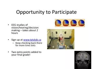
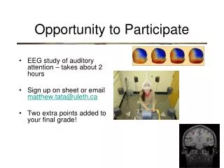
Opportunity to Participate • EEG study of auditory attention – takes about 2 hours • Sign up on sheet or email matthew.tata@uleth.ca • Two extra points added to your final grade!
Functional Imaging • blood flow overshoots baseline after a brain region is activated • More oxygenated blood in that region increases MR signal from that region (other regions de-phase faster)
Functional Imaging • It is important to recognize that fMRI “sees” changes in the ratio of oxygenated to deoxygenated blood - nothing more • BOLD: Blood Oxygenation Level Dependant contrast • How do we create those pretty pictures?
Functional Imaging • It is important to recognize that fMRI “sees” changes in the ratio of oxygenated to deoxygenated blood - nothing more • BOLD: Blood Oxygenation Level Dependant contrast • How do we create those pretty pictures? • We ask the question “When the subject engages in this cognitive task, where does blood oxygenation change significantly?” “where does it change randomly?”
Experimental Design in fMRI • Experimental Design is crucial in using fMRI • Simplest design is called “Blocked” • alternates between active and “rest” conditions Active Rest Active Rest 60 sec 60 sec 60 sec 60 sec
Experimental Design in fMRI • Experimental Design is crucial in using fMRI • Simplest design is called “Blocked” • alternates between active and “rest” conditions Active Rest Active Rest 60 sec 60 sec 60 sec 60 sec
Experimental Design in fMRI • A voxel in tissue insensitive to the task demands shows random signal change Signal Active Rest Active Rest 60 sec 60 sec 60 sec 60 sec
Experimental Design in fMRI • A voxel in tissue that responds to the task shows signal change that matches the timecourse of the stimulus Signal Active Rest Active Rest 60 sec 60 sec 60 sec 60 sec
Experimental Design in fMRI • A real example of fMRI design done well: • alternate moving, blank and stationary visual input Moving Blank Stationary Blank 40 sec 40 sec 40 sec 40 sec
Experimental Design in fMRI • Voxels in Primary cortex tracked all stimuli
Experimental Design in fMRI • Voxels in area MT tracked only the onset of motion
Experimental Design in fMRI • Voxels in area MT tracked only the onset of motion • How did they know to look in area MT?
PET: another way to measure blood Oxygenation • Positron Emission Tomography (PET) • Injects a radioisotope of oxygen • PET scanner detects the concentration of this isotope as it decays
PET: another way to measure blood Oxygenation • Although oxygenation is measured differently, the logic of PET and fMRI are similar: compare active and “rest” conditions
Advantages of fMRI • All techniques have certain advantages • A good scientist leverages these advantages
Advantages of fMRI • Advantages of MRI: • Most hospitals have MRI scanners that can be used for fMRI (PET is rare) • Better spatial resolution in fMRI than PET • Structural MRI is usually needed anyway • No radioactivity in MRI • Better temporal resolution in MRI
Advantages of PET • Advantages of PET: • Quiet • A number of different molecules can be labeled and imaged in the body
Limitations of fMRI • All techniques have constraints and limitations • A good scientist is careful to interpret data within those constraints
Limitations of fMRI • Limitations of MRI and PET: • BOLD signal change does not necessarily mean a region was specifically engaged in a cognitive operation • Poor temporal resolution - depends on slow changes in blood flow • expensive