The Head and Face
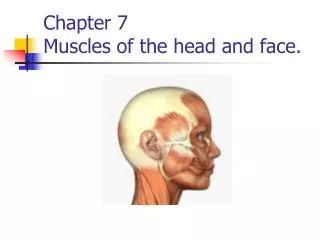
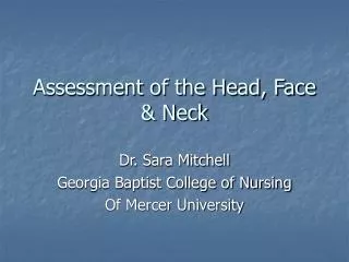
The Head and Face. Chapter 23. The Head and Face. KEY CONCEPT The head can be divided into two anatomical groups; the face and the cranium. The face includes the structures of the eye, nose, mouth, ears, and jaw. The cranium includes the brain, skull, and spinal cord attachments.

The Head and Face
E N D
Presentation Transcript
The Head and Face Chapter 23
The Head and Face • KEY CONCEPT The head can be divided into two anatomical groups; the face and the cranium. The face includes the structures of the eye, nose, mouth, ears, and jaw. The cranium includes the brain, skull, and spinal cord attachments.
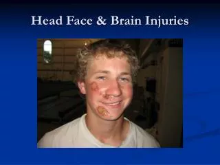
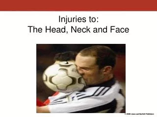
The Head and Face • The head can be divided into two anatomical groups: the face and the cranium. • The face includes the eyes, ears, nose, jaw, and mouth. • The cranium includes the brain and spinal cord attachments. • The term head injury may be used to describe damage to the scalp, skull, or brain, usually as the result of the application of a sudden force to the head.
The Eye • The eye is protected in many ways from the outside world. • Eyebrows, eyelashes, and eyelids provide physical barriers to objects that might get into the eye. • Tears, produced by nearby lacrimal glands, keep the eye moist, wash away small foreign objects that make it into the eye, and contain an enzyme capable of destroying bacteria. • Glands along the border of each eyelid secrete a substance that helps smooth the surface and lubricate the eye. An infection of these glands is called a sty. • The conjunctiva is a thin membrane that covers the entire front of the eye and part of the inner eyelid.
The Eye • The sclera, or white of the eye, is a thick, fibrous layer that maintains the shape of the eye and protects the structures within. It is attached to the extrinsic eye muscles that are responsible for the eye movements.
The Eye • The cornea is continuous with the sclera, but is transparent, allowing light to pass through into the inner structures of the eye. Its convex shape works along with the lens to create a sharp image on the retina of the eye.
The Eye • The choroid coat lines the inner sclera and is darkly pigmented to prevent random reflections of light entering the eye’s inner structures. • The choroid coat is continuous with the iris-a variably pigmented layer that has a central opening called the pupil, through which light passes to get to the eye’s internal structures. • The intrinsic eye muscles are located within the iris, allowing it to change the size of the pupil and respond to the amount of light available.
The Eye • The lens is transparent, crystalline structure attached by suspensory ligaments to the ciliary body that controls the thickness of the lens. • As the lens increases in thickness, the eye is able to sharply focus nearby objects on to the retina. • The anterior chamber and the posterior chamber are filled with a watery fluid called the aqueous humor. • Behind the lens is a area filled with a jellylike substance, called the vitreous humor, that extends back to the retina and helps maintain the shape of the eye.
Lens, Suspensory Ligaments, Ciliary Body, Aqueous Humor, and Vitreous Humor
The Eye • The retina is the thin layer of cells located between the vitreous humor and the choroid coat that contains the light-sensitive cells called rods and cones. • The rods are sensitive to dim light; since there is only one type of rod, they cannot distinguish colors. • The cones are active in bright light. There are three different kinds of cones, each sensitive to a different range of light frequencies, that allow for color vision. • The fovea centralis is an area of the retina especially rich in cones, where the retina is the thinnest, providing the sharpest vision. The other parts of the retina provide our peripheral vision, which is not as sharp. • The optic disc is located where the nerve fibers from the rods and cones leave the eye and enter the optic nerve. The optic disc has no rods or cones, so it is calso called the blind spot.
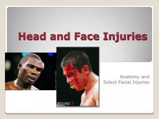
Eye Injuries • Key Concept • Dirt and debris can become imbedded into the structures of the eye and cause pain or a corneal abrasion. A corneal abrasion is a cut or scratch on the cornea. Attempts can be made to wash objects out of the eye by splashing water into the eye. However, imbedded objects should not be removed and the athlete should be instructed to see a physician. • A blow or contusion to the eye often results in a black eye. Application of a cold compress for 15 minutes immediately after the injury will aid in reducing pain and swelling. • Cuts, punctures, or abrasions to the eye or eyelid can cause infection and blindness. These are medical emergencies and the athlete should be promptly transported to a medical facility. • An orbital blow-out fracture is a break in the bones that house the eye. Immediate treatment consists of bandaging both eyes and applying an ice compress for 15 to 20 minutes. The athlete should seek immediate treatment from an ophthalmologist. • Hyphemia is the buildup of blood in the anterior chamber of the eye. Treatment will depend on the cause, but the athlete should seek the care of an ophthalmologist. • Conjunctivitis is an infection of the outermost layer of the eye. This is usually contagious and requires medical attention.
Eye Injuries • Specks in the eyes can cause a corneal abrasion, scratches, or cuts on the cornea. • Objects should be washed out by splashing clean water into the eye. • If the object cannot be removed, or is imbedded in the eye, the athlete should see a doctor immediately.
Eye Injuries • Blows (contusions) to the eye are common in sports. The eye is located in a deep socket call the orbit. • Symptoms include pain, swelling, and discoloration. • Treatment includes applying a cold compress immediately for 15 minutes, and again each hour as needed. If there is discoloration or blackening of the eye, the athlete should consult a physician immediately.
Eye Injuries • Cuts, punctures, and abrasions of the eye or eyelid are medical emergencies and require prompt transport to the nearest medical facility.
Eye Injuries • An orbital blow-out fracture consists of a fracture of the bones of the eye socket and is usually secondary to a blunt blow from a relatively large object. • Symptoms include pain, tenderness, swelling, bruising, double vision (diplopia), protrusion of the eye (proptosis), and/or numbness in the cheek and upper jaw areas. • Bandage both eyes and apply an ice compress for 15 to 20 minutes. The athlete should be sent to an ophthalmologist.
Eye Injuries • Hypheme refers to bleeding in the anterior chamber of the eye, due to bleeding of the vessels of the iris. • Symptoms include the athlete complaining of dramatically decreased vision. • Athletes with hypheme should be seen by an ophthalmologist, even though the blood is often reabsorbed over a period of days to weeks.
Eye Injuries • Conjunctivitis, or pinkeye, is an infection of the conjunctiva, which can be caused by a virus, bacteria, or an allergic reaction. • The viral and bacterial forms are typically contagious. • Symptoms include eye discomfort followed by redness and inflammation of the conjunctiva. After a day or so a white, yellow, or green discharge form the eyes may be present. • Treatment requires medical attention and depends on the cause.
The Ear • The ear consists of three parts: the outer ear, the middle ear, and the inner ear. • The outer ear is composed of the pinna and the ear canal. • The pinna (auricle) is the visible part of the ear composed of folds of skin and cartilage. • The ear canal (also called the meatus) is a short tube leading to the tympanic membrane (eardrum). It produces wax that, along with tiny hairs in the canal help trap dust and small foreign bodies.
The Ear • The middle ear is an air-filled space between the eardrum and the inner ear. Its hearing structures consist of three small bones called ossicles. It contains the eardrum, hammer, anvil, stirrup, and eustachian tube. • The malleus (hammer) is attached to the inside of the eardrum. • The incus (anvil) connects the malleus to the stapes. • The stapes (stirrup) is the third bone, which attaches the incus to the oval window of the inner ear. • The eustachian tube connects the middle ear to ehe throat. It is closed unless the person is swallowing or yawning. It then opens the space of the middle ear to outside air, equalizing air pressure in the middle ear to that of the outside.
The Ear • The inner ear consists of an extremely intricate series of structures contained within the bones of the skull. • The cochlea is a coiled tube containing the sensory nerves for the sense of hearing. • The semicicular canals contain the sensory nerves for detecting how the head is moving. • A cavity known as thevestibulecontains the sensory nerves that inform the brain about the current position of the head. • These structures are connected to the vestibulocochlear nerve, which carries the sensory information to the nearby brain.
Injuries to the Ear • Cauliflower ear is caused by the destruction of the underlying cartilage of the outer ear (pinna). Blood collects between the cartilage and skin, causing a thickening of the entire outer ear. • Symptoms include a blood clot under the skin of the ear, which will eventually cause the ear cartilage to die and shrivel up, since the skin is its only blood supply. • Treatment of the hematoma (blood clot) is to drain it through an incision in the ear and apply a compressive dressing to sandwich the two sides of skin against the cartilage.
Injuries to the Ear • Swimmer’s ear is an infection of the skin covering the outer ear canal. The ear needs to be kept dry, since moisture will irritate and prolong the problem. • Foreign bodies in the ear, including insects, can be difficult to remove because of the small size of the ear canal. • Symptoms include mild to severe ear pain, drainage from the ear, fever, nausea and vomiting, coughing, tearing from the eye, dizziness, and a foul odor from the ear caused by infection. • Treatment involves anything from gentle flushing of the ear canal with warm water to surgery to remove the object.
Injuries to the Ear • Tympanic rupture is most often caused by a middle ear infection, but may be due to trauma. • Symptoms include severe ear pain and sudden drainage, if caused by an infection. • Treatment, whatever the cause, involves immediate transport to a physician for evaluation and treatment.
The Nose • The outer nose is composed of bone, cartilage, and skin, and projects from the front of the face, making it susceptible to injury. • The human nose serves as an air passage for the respiratory system and provides the brain with the sense of smell.
Injuries of the Nose • Epistaxis is the medical term for nosebleed, though it usually refers to recurrent nosebleeds or those difficult to stop. • A sign of posterior epistaxis is an athlete complaining of swallowing blood. There is no way for the certified athletic trainer to stop this type of nosebleed. Posterior nosebleeds can be life-threatening and should be considered a medical emergency. • For anterior epistaxis, the athlete should sit and lean slightly forward. The athlete should squeeze the soft portion of the nose for about 5 minutes and repeat if necessary. A cold compress across the bridge of the nose can help.
Injuries to the Nose • Nasal fractures and septal deviations occur as the result of direct blows or as the result of falls. The nasal bones are the most commonly fractured bony structures of the face. • Symptoms include signs of deformity, swelling, skin laceration, ecchymosis, epistaxis, and leakage of cerebrospinal fluid (CSF). • Treatment begins with careful, direct pressure, the application of ice, and getting the athlete to sit with the head tilted slightly forward. The athlete should be sent to a physician for additional care and treatment.
The Mouth and Jaw • The mouth includes structures like the soft palate and hard palate, mucous membranes, tongue, teeth, lips and cheeks. • The upper jaw includes the maxilla bone, which is fixed to the skull. • The lower jaw is the mandible bone, which is attached at a moveable joint on the temporal bone of the skull, called the temporomandibular joint (TMJ).
Injuries to the Mouth and Jaw • Jaw fractures usually include two fractures, one direct, and one indirect. The indirect fracture is usually located near one of the condyles of the mandible close to the joint. • Symptoms include severe pain, swelling, blood at he base of the teeth near the fracture, deformity, tenderness, and sometimes numbness. • Treatment includes immobilization, application of ice, and treatment for shock. The athlete should be transported to a physician immediately.