Download
1 / 1
10 likes | 195 Vues
P lasma treatment of caries : a novel method in dentistry. R.E.J. Sladek, R. Walraven, E. Stoffels, P.J.A. Tielbeek, R.A. Koolhoven Department of Biomedical Engineering , Eindhoven University of Technology , P.O. Box 513, 5600 MB Eindhoven, The Netherlands E-mail: R.E.J.Sladek@tue.nl.

E N D
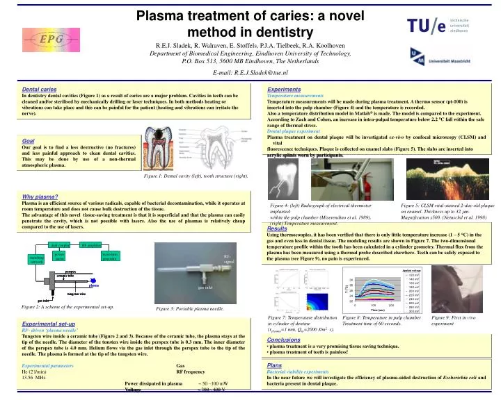
Plasma treatment of caries: a novel method in dentistry R.E.J. Sladek, R. Walraven, E. Stoffels, P.J.A. Tielbeek, R.A. KoolhovenDepartment of Biomedical Engineering, Eindhoven University of Technology, P.O. Box 513, 5600 MB Eindhoven, The Netherlands E-mail: R.E.J.Sladek@tue.nl Dental caries In dentistry dental cavities (Figure 1) as a result of caries are a major problem. Cavities in teeth can be cleaned and/or sterilised by mechanically drilling or laser techniques. In both methods heating or vibrations can take place and this can be painful for the patient (heating and vibrations can irritate the nerve). Experiments Temperature measurements Temperature measurements will be made during plasma treatment. A thermo sensor (pt-100) is inserted into the pulp chamber (Figure 4) and the temperature is recorded. Also a temperature distribution model in Matlab® is made. The model is compared to the experiment. According to Zach and Cohen, an increase in intra-pulpal temperature below 2.2 C fall within the safe range of thermal stress. Dental plaque experiment Plasma treatment on dental plaque will be investigated ex-vivo by confocal microscopy (CLSM) and vital fluorescence techniques. Plaque is collected on enamel slabs (Figure 5). The slabs are inserted into acrylic splints worn by participants. Goal Our goal is to find a less destructive (no fractures) and less painful approach to clean dental cavities. This may be done by use of a non-thermal atmospheric plasma. Figure 1: Dental cavity (left), tooth structure (right). Why plasma? Plasma is an efficient source of various radicals, capable of bacterial decontamination, while it operates at room temperature and does not cause bulk destruction of the tissue. The advantage of this novel tissue-saving treatment is that it is superficial and that the plasma can easily penetrate the cavity, which is not possible with lasers. Also the use of plasmas is relatively cheap compared to the use of lasers. Figure 4: (left) Radiograph of electrical thermistor implanted within the pulp chamber (Miserendino et al. 1989). (right) Temperature measurement. Figure 5: CLSM vital-stained 2-day-old plaque on enamel. Thickness up to 32 m. Magnification x500. (Netuschil et al. 1998) Results Using thermocouples, it has been verified that there is only little temperature increase (1 – 5 °C) in the gas and even less in dental tissue. The modeling results are shown in Figure 7. The two-dimensional temperature profile within the tooth has been calculated in a cylinder geometry. Thermal flux from the plasma has been measured using a thermal probe described elsewhere. Teeth can be safely exposed to the plasma (see Figure 9), no pain is experienced. Figure 2: A scheme of the experimental set-up. Figure 3: Portable plasma needle. Figure 7: Temperature distribution in cylinder of dentine (rplasma=1 mm, Qin=2000 J/m2 . s). Figure 8: Temperature in pulp chamber Treatment time of 60 seconds. Figure 9: First in vivo experiment Experimental set-up RF- driven ‘plasma needle’ Tungsten wire inside a ceramic tube (Figure 2 and 3).Because of the ceramic tube, the plasma stays at the tip of the needle. The diameter of the tunsten wire inside the perspex tube is 0.3 mm. The inner diameter of the perspex tube is 4.0 mm.Helium flows via the gas inlet through the perspex tube to the tip of the needle. The plasma is formed at the tip of the tungsten wire. Experimental parametersGas He (2 l/min)RF frequency 13.56MHz Power dissipated in plasma 50 –100 mW Voltage 200 - 400 V • Conclusions • plasma treatment is a very promising tissue saving technique. • plasma treatment of teeth is painless! Plans Bacterial viability experiments In the near future we will investigate the efficiency of plasma-aided destruction of Escherichia coli and bacteria present in dental plaque.