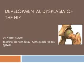
Hip Dysplasia
Hip Dysplasia. Diagnosis and Classification Schemes Erica Fields, DVM. Cause(s) of Hip Dysplasia. Multifactorial Heritable disorder Nutritional factors (overnutrition) Early exercise types/amounts Even season of birth has been shown to affect expression. Not Just Dogs….

Hip Dysplasia
E N D
Presentation Transcript
Hip Dysplasia Diagnosis and Classification Schemes Erica Fields, DVM
Cause(s) of Hip Dysplasia • Multifactorial • Heritable disorder • Nutritional factors (overnutrition) • Early exercise types/amounts • Even season of birth has been shown to affect expression Thrall, Textbook of Vet Rad and Ohlerth, et al. 2000.
Not Just Dogs… • Yes, we usually associate hip dysplasia with large, working-breed dogs, BUT • Cats and small or toy breed dogs can also be affected • Incidence in DSH estimated at 6.6%, Maine Coons up to 21% • Some large breed dogs more susceptible than others (GSD vs. Greyhound) Thrall. 4th ed Textbook of Vet Rad. 2002.
Signs of Hip Dysplasia • Physical examination • “Walking Ortolani”—hands on trochanters during normal walk—palpate for laxity • Ortolani sign—dorsal recumbency, abduct hips, feel for “clunk” Young dogs • Barden’s Hip Lift—lateral recumbency, anesthetized, lift hip laterally. Greater than 5-6 mm movement=positive • Gait abnormalities (swing/bunny hop) WSAVA Congress Proceedings 2002 and 2005
Signs of Hip Dysplasia (cont’d) • Radiographic signs • Joint laxity (distraction index, subluxation) • Osteoarthritic changes • Enthesophytosis at insertion of joint capsule on caudal aspect of femoral neck (Morgan line) • Osteophytes • Femoral head/neck remodeling • Acetabular remodeling • Subchondral sclerosis of femoral head/acetabulum Thrall, 4th ed. Textbook of Vet Rad
What about cats? • Coxofemoral subluxation • Most degenerative changes appear on the craniodorsal acetabulum, not the femoral head and neck Thrall 4th ed Textbook of Vet Rad
Radiographic Detection of Hip Dysplasia • MANY methods—each has its advantages • “OFA” view—extended hip VD • DAR view—better visualization of acetabular rim blunting • Ultrasonographic evaluation • PennHIP distraction method • European (Federation Cynologique International)—five point quality scale numerical system based on 6 factors • British (BVA) system— • German system
OFA Classifications • Extended hip VD pelvis view • Consensus of 3 boarded radiologists • Based on evaluation of 9 anatomic areas (craniolateral acetabular rim, cranial acetabular margin, femoral head, fovea capitis, acetabular notch, caudal acetabular rim, dorsal acetabular margin, femoral head/neck junction, trochlear fossa)
OFA guidelines (cont’d) • Radiographs must be performed after 24 mos of age—based on a study showing that only 16% of dysplastic GSDs were diagnosed at 6 mos, as compared to 95% at 24 mos • Six classes—1 through 6 (excellent, good, fair, borderline, mild, moderate, severe) Ohlerth, et al. J Sm An Pract 2003.
Pros and Cons—OFA • Pro • Easy to perform, no special tools or certifications needed • Good identification of osteoarthritic changes • Large centralized database • Well-recognized and established in the breeding community • Compares dogs within breeds
Pros and Cons—OFA • Cons • Extended hip view may artificially tighten joint • Not a physiologic position • Selection bias (really bad ones don’t get submitted) • Final eval can’t be done before 24 months—delays breeding times • Little eval of laxity
DAR view • Can supplement other views to better evaluate changes in dorsal acetabular rim • Used in planning for TPO procedures • Blunting of DAR caused by microfractures due to altered load bearing WSAVA World Congress 2002
Ultrasound evaluation • Accepted method in infants • In development in dogs • Done with a distraction device in place, longitudinal view in inguinal region—dynamic study • Difficult to perform accurately, variation may alter measurements • Only appears to be reliable in predicting true negatives Ohlerth et al. J Sm An Pract. 2003
PennHIP • Three views—standard OFA view, compression view with legs bent at 90 degrees, distraction view with legs bent at 90 degrees • All radiographs are submitted to central database • Distraction index calculated based on hip geometry • Distraction indices correlated to likelihood of OA development for various breeds
PennHIP—Pros and Cons • Pros • Can be performed as early as 4-5 mos, resulting in better breeder screening • Anatomic positioning—more functional evaluation and less artificial joint tightening • Less selection bias • Evaluates laxity, an early cause/indicator of CHD Thrall 4th ed Textbook of Vet Rad
PennHIP—Pros and Cons • Cons • Specialized device and training necessary • Must submit all radiographs To get around these disadvantages, some people have tried to develop other techniques, including using wooden distractors or performing Ortolani and calculating subluxation Thrall 4th ed and Ohlerth, et al 2003
Federation Cynologique Internationale System (FCI) • Uses 6 criteria that are scored 0-5 (0 good, 5 bad) • Norberg angle (more on this later) • Coverage of femoral head by acetabular rim • Craniodorsal acetabular rim conformation/osteophytosis • Subchondral bone sclerosis • Femoral head/neck shape and osteophytosis • Joint capsule insertion enthesophytosis Ohlerth et al. AJVR 2001
British Veterinary Association • 12 mos minimum age; no maximum • No resubmissions (unlike OFA, where prelim eval is allowed) • VD extended hip (OFA) view • Nine criteria evaluated and scored 0-6 PER HIP. Scores for hips are added for a total score—best score is 0/worst is 53 for each hip, or 106 total score. • Breed mean scores are published www.bva.co.uk/public
BVA Criteria • Norberg angle • Subluxation • Cranial acetabular edge • Dorsal acetabular edge • Cranial effective acetabular rim • Acetabular fossa • Caudal acetabular edge • Femoral head/neck osteophytosis • Femoral head remodeling www.bva.co.uk/public
Comparison of Different Scoring Systems OFA FCI (Euro) BVA (Aust/UK) SV (Ger) E A-1 0-4 (no >3/hip) Normal G A-2 5-10 (no >6/hip) Normal F B-1 11-18 Normal B B-2 19-25 Fast Norm M C 26-35 Noch Zugelassen Mod D 36-50 Mittlere S E 51-106 Schwere www.offa.org
Norberg Angle • Used in many evaluation strategies • Evaluation of cranial acetabular morphology and subluxation • A line is drawn between center points of both femoral heads and lines drawn from each femoral head center to the craniolateral aspect of DAR • Angle is calculated between 2 lines Tomlinson et al AJVR 2000
Norberg Angle • Traditionally, Norberg angle of greater than 105 degrees and acetabular coverage of greater than 50% is considered normal • A study of 4 most common breeds in OFA registry compared NA, coverage, and OFA classification • The 105/50 rule does not hold constant across breeds. • Norberg angles from 92.6 (Goldens) to 101.9 (Rotties) are correlated with normal OFA Tomlinson et al. AJVR 2000
References • Allan G.: Radiographic signs of joint disease. In Thrall, D (ed): Textbook of Veterinary Radiology, 4th ed. Philadelphia. WB Saunders Co, 2002. pp 190-195. • Ohlerth S, Busato A, Rauch M, Weber U, Lang J. Comparison of three distraction methods and conventional radiography for early diagnosis of canine hip dysplasia. Journal of Small Animal Practice. 2003 44:524-529. • Ohlerth S, Lang J, Busato A, Gaillard C. Estimation of genetic population variables for six radiographic criteria of hip dysplasia in a colony of Labrador Retrievers. AJVR 2001 62(6): 846-852. • Tomlinson JL, Johnson JC. Quantification of measurement of femoral head coverage and Norberg angle within and among four breeds of dogs. AJVR 2000 61(12): 1492-1500. • www.bva.co.uk/public • www.offa.org