Download
1 / 186
1.86k likes | 1.96k Vues
Discover the essential components of the peripheral nervous system, including sensory receptors, nerves, and ganglia. Learn how sensory receptors detect stimuli and transmit signals to the CNS through afferent fibers. Explore the classification of nerves and ganglia based on their functions and structure.

E N D
The Peripheral Nervous System Chapter 14
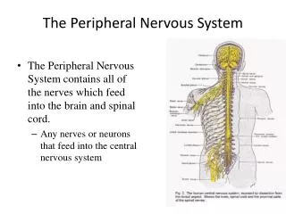
Introduction • The CNS would be useless without a means of sensing our own internal as well as the external environments • In addition, we need a means by which we can effect our external environment • The peripheral nervous system provides these links to the CNS
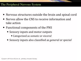
Introduction • The peripheral nervous system includes all the neural structures outside the brain and spinal cord • Sensory receptors • Peripheral nerves and their ganglia • Efferent motor endings
Introduction • Basic components of the PNS • Sensory components provide the information interpreted by the CNS • Motor components stimulate the effectors of the CNS • The CNS commands; the PNS acts
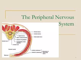
Nerves and Associated Ganglia • A nerve is a cordlike organ that is part of the peripheral nervous system • Every nerve consists of parallel bundles of peripheral axons enclosed by successive wrappings of connective tissue
Nerves and Associated Ganglia • Within a nerve, each axon is surrounded by a delicate layer of loose connective tissue called endoneurium • The endoneurium layer also encloses the fiber’s associated myelin sheath
Nerves and Associated Ganglia • Groups of fibers are bound into bundles or fascicles by a courser connective tissue wrapping called the perineurium • All the fascicles are enclosed by a tough fibrous sheath called the epineurium to form a nerve
Nerves and Associated Ganglia • Neurons are actually only a small fraction of the nerve • The balance is myelin, the protective connective tissue wrappings, blood vessels, and lymphatic vessels
Nerves and Associated Ganglia • Nerves are classified according to the direction in which they transmit impulses • Nerves containing both sensory and motor fibers are called mixed nerves • Nerves that carry impulses toward the CNS only are sensory (afferent) nerves • Nerves that carry impulses only away from the CNS are motor (efferent) nerves • Most nerves are mixed as purely sensory or motor nerves are extremely rare
Nerves and Associated Ganglia • Since mixed nerves often carry both somatic and autonomic (visceral) nervous system fibers, the fibers within them may be classified further according to the region they innervate as • Somatic afferent • Somatic efferent • Visceral afferent • Visceral efferent
Nerves and Associated Ganglia • Peripheral nerves are generally classified on whether they arise from the brain or spinal cord as • Cranial nerves / brain and brain stem • Spinal nerves / spinal cord • Ganglia are collections of neuron cell bodies associated with nerves in the PNS • Ganglia associated with afferent nerve fibers contain cell bodies of sensory neurons • Ganglia associated with efferent nerve fibers contain cell bodies of autonomic neurons, as well as a variety of integrative neurons
Sensory Receptors • Sensory receptors are structures that are specialized to respond to changes in their environment • Such environmental changes are called stimuli • Typically activation of a sensory receptor by an adequate stimulus results in depolarization or graded potentials that trigger nerve impulses along the afferent fibers coursing to the CNS
Peripheral Sensory Receptors • Peripheral sensory receptors are structures that pick up sensory stimuli and then initiate signals in the sensory axons • Most receptors fit into two main categories; • Dendritic endings of sensory neurons • Complete receptor cells
Peripheral Sensory Receptors • Dendritic endings of sensory neurons monitor most types of general sensory information (touch, pain, pressure, temperature, and proprioception)
Peripheral Sensory Receptors • Complete receptor cells are specialized epithelial cells or small neurons that transfer sensory information to sensory neurons • Specialized receptor cells monitor most types of special sensory information (taste, vision, hearing, and equilibrium)
Sensory Receptors • Sensory receptors are classified by • The type of stimulus they detect • Their location in the body • Their structure
Classification by Location • Receptors are recognized according to their location or the location of the stimuli to which they respond • Externoceptors • Internoceptors or visceroceptors • Proprioceptors
Classification by Location • Externoceptors • Sensitive to stimuli arising from outside of the body • Typically located near the surface of the body • Include receptors for • Touch • Pressure • Pain • Temperature • Special sense receptors
Classification by Location • Internoceptors or visceroceptors • Respond to stimuli arising from within the internal viscera and body organs, • Internoceptors monitor a variety of internal stimuli • Changes in chemical concentration • Taste stimuli • The stretching of tissues • Temperature • Their activation causes us to feel visceral pain, nausea, hunger, or fullness
Classification by Location • Proprioceptors • Located in the musculoskeletal organs such as skeletal muscles, tendons, joints and ligaments • Proprioceptors monitor the degree of stretch of these locomotor organs and send input to the CNS
Classification by Stimulus Detected • Mechanoreceptors • general nerve impulses when they, or adjacent tissues, are deformed by mechanical forces • Touch • Pressure • Vibration • Stretch • Itch • Thermoreceptors • Sensitive to temperature changes
Classification by Stimulus Detected • Photoreceptors • Respond to light energy • Chemoreceptors • Respond to chemicals in solution • Smell • Taste • Blood chemistry • Nociceptors • Respond to potentially damaging stimuli that result in pain
Classification by Stimulus Detected • Note that the over-stimulation of any of the aforementioned receptors is painful and thus virtually all receptors can function as nociceptors at one time or another
Classification by Structure • General sensory receptors are divided into two broad groups • Free (naked) endings • Encapsulated dendritic endings • It should be pointed out that there is no one receptor - one function relationship • Rather, one receptor type can respond to several different kinds of stimuli, and different receptor types can respond to similar stimuli
Adaptation of Sensory Receptors • Adaptation occurs in certain sensory receptors when they are subjected to an unchanging stimulus • As a result, the receptor potentials decline in frequency or stop • Some receptors adapt quickly (pressure, touch and smell) • Nocioceptors and proprioceptors adapt slowly or not at all as they serve a protective function
Free Dendritic Endings • Free nerve endings have small knoblike swellings • Chiefly respond to pain, temperature, and possible mechanical pressure caused by tissue movement
Free Dendritic Endings • The receptors are simple and widely dispersed everywhere in the body • Particularly abundant in epithelia and connective tissue underlying epithelial tissue
Merkel Discs • Certain free dendritic endings contribute to Merkel discs • These discs lie in the epidermis of the skin
Merkel Cells • Merkel cells attach to the basal layer of the skin epidermis • Each Merkel disc consists of a disc- shaped epithelial cell innervated by a dendrite • Functions as light touch receptors
Merkel Discs • Merkel cells seem to be slowly adapting receptors for light touch • Slowly adapting means that they continue to respond to stimuli present and send out action potentials even long after a period of continual stimulation
Root Hair Plexuses • Root hair plexuses are free dendritic endings that wrap around hair follicles • These are receptors for light touch that monitor the bending of hairs
Root Hair Plexuses • Root hair plexuses are rapidly adapting • This means that the sensation disappears quickly even if the stimulus is maintained • The landing of a mosquito is mediated by root hair plexuses Root Hair Plexus
Encapsulated Dendritic Endings • All encapsulated dendritic endings consist of one or more end fibers of sensory neurons enclosed in a capsule of connective tissue • All seem to be mechanoreceptors, and their capsules serve to either amplify the stimulus or to filter out the wrong types of stimuli
Encapsulated Dendritic Endings • Encapsulated receptors vary widely in shape, size, and distribution in the body • The main types are • Meissner’s corpuscles • Krause’s end bulbs • Pacinian corpuscles • Ruffini’s corpuscles • Proprioceptors
Meissner’s Corpuscles • In a Meissner’s corpuscle (tactile corpuscle) a few spiraling dendrites are surrounded by Schwann cells, which in turn are surrounded by an egg-shaped capsule of connective tissue
Meissner’s Corpuscles • These corpuscles are found in the dermal papillae beneath the epidermis • These corpuscles are rapidly adapting receptors for fine, light touch
Meissner’s Corpuscles • Meissner’s corpuscles occur in sensitive and hairless areas of the skin, such as the soles of the feet, palms, fingertips, nipples, and lips • Apparently, Meissner’s corpuscles perform the same “light touch” function in hairless skill that root hair plexuses perform in hairy skin
Krause’s End Bulbs • Krause’s End Bulbs are a type of Meissner’s corpuscle for fine touch • Krause’s end bulbs occur in mucous membranes in the lining of the mouth and the conjunctiva of the eye
Pacinian Corpuscle • Pacinian corpuscle are scattered throughout the deep connective tissues of the body • Occur in the hypodermis of the skin
Pacinian Corpuscles • Pacinian corpuscles contains a single dendrite surrounded by up to 60 layers of Schwann cells and is in turn enclosed by connective tissue • Respond to deep pressure • Rapidly adapting as they respond to only the initial pressure
Pacinian Corpuscles • Pacinian corpuscles are rapidly adapting receptors and are best suited to monitor vibrations which is an on-off stimulus • These corpuscles are large enough to be visible to the naked eye
Ruffini’s Corpuscle • Ruffini’s corpuscle are located in the dermis of the skin and joint capsules of the body • The corpuscle contains an array of dendritic endings enclosed in a thin flattened capsule
Ruffini’s Corpuscle • Ruffini’s corpuscle respond to pressure and touch • They adapt slowly and thus can monitor continuous pressure placed on the skin
Virtually all proprioceptors are encapsulated dendritic endings that monitor stretch in the locomotor organs Proprioceptors include… Muscle spindles Golgi tendon organs Joint kinesthetic receptors Proprioceptors
Proprioceptors • Muscle spindles measure the changing length of a muscle as that muscle contracts and as it is stretched back to its original length • Muscle spindles are found throughout skeletal muscle
Proprioceptors • An average muscle contains some 50 to 100 muscle spindles, which are embedded in the perimysium between muscle fascicles
Muscle Spindles • Structurally each muscle spindle consists of a bundle of modified skeletal muscle fibers called intrafusal fibers enclosed in a connective tissue capsule • Infrafusal fibers have fewer striations than do the ordinary muscle cells
Proprioceptors • The intrafusal fibers are innervated by the dendrites of several sensory neurons