Case Report
Case Report. DISIDA Scan. Case I. Name: 劉亦承 Age: 2 m/o Sex: Male. Chief complain. Prolonged jaundice since birth . Present illness. A case of full-term(BBW:3400gm;NSD) born Jaundice was noted since he was 3-4 d/o. After going home, pale looking and jaundice was still noted.


Case Report
E N D
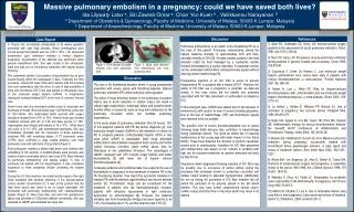
Presentation Transcript
Case Report DISIDA Scan
Case I • Name: 劉亦承 • Age: 2 m/o • Sex: Male
Chief complain Prolonged jaundice since birth
Present illness • A case of full-term(BBW:3400gm;NSD) born • Jaundice was noted since he was 3-4 d/o. • After going home, pale looking and jaundice was still noted. • Impaired liver function (GOT/GPT:76/50) and hyperbilirubinemia (Bil(T/D):4.6/2.7) • Abdominal echo revealed hepatomegaly.
Physical examination • Cons: alert; conj: pale; sclera: not icteric • HEENT: np • Neck: supple, LAP(-) • Chest: symmetric expansion • HS:RHB, no murmur • BS: no rale • Abdominal: soft, ovoid, • BS: normoactive ; Liver :2fb below RCM • Limbs: free movable
Differential Diagnosis • Common: 1. Biliary atresia 2. Neonatal hepatitis • Uncommon: 1. Sepsis 2. Infectious hepatitis(TORCH, syphilis, HBV, Rubella) 3. Alpha-1 Antitrypsin Deficiency) 4. Other causes for high-grade obstruction
Plan • HbsAg • AFP • TORCH + VDRL, α1-antitrypsin • CMV (urine):- ; CMV (serum): + • Rub IgG: + • Liver biopsy • Ultrasound • DISIDA
Liver Biopsy • Fatty metamorphosis, cholestasis and the presence of giant cells transformation. • Portal areas are still shown, without bile ductules proliferation.
Abdominal Echo • Liver: homogenous parenchyma and enlarged size • GB and bile duct: normal • Portal vein and hepatic vein: normal • Pancreases and spleen: normal • Ascites:Nil; no occupy lesion is noted. IMP: Hepatomegaly
Indication • Evaluation cholecysitis • Differentiate between obstructive and nonobstructive jaundice • Investigate persons with upper abdominal pain • Biliary assessment postsurgery and evaluation of biliary atresia
Does not have true hepatocellular dysfunction such that tracer accumulation in the liver should be normal. Bowel activity should not been seen. Abnormal liver function leading to depressed hepatocellular accumulation of tracer and delayed transit through the liver. Bowel activity will eventually been seen Biliary atresia&Neonatal hepatitis
Definite Diagnosis Neonatal hepatitis
Case II • Name: 呂彥霖 • Age: 1m 4d/o • Sex: Male
Chief Compliance Prolonged jaundice since birth
Present Illness • This 1m 4d/o male infant was born on 90-04-14. • Prolonged jaundice was found later. • He was brought to LMD and then was referred to our CNU due to elevated bilirubin level. (Bil(T/D):5.6/1.8)
Physical Examination • Cons: alert; sclera: mild icteric • Neck: supple, LAP (-) • Chest: symmetric expansion • HS: RHB, no murmur • BS: clear • Abd: Liver: 2cm below RCM • Spleen: just palpable • Limbs: free movable ; No pitting edema
Tc 99m DISIDA cholescintigraphy • Good hepatic uptake on the 5 min image • Homogeneous radiotracer distribution of liver on the 15 min image. • Visualized radioactivity of GB on the 30-min image. • Relatively low amount of intestinal radioactivity on the serial one and six hours images. • Persistent hepatic radioactivity on the 24 hour image.
Impression • Impaired bile passage, partial biliary atresia is considered. • Functional GB.
MRCP • Revealed visible common bile duct • The gall bladder was roughly 3 cm in length • Intrahepatic ducts were not dilated • No abnormal signal intensity lesion in the liver parenchyma. • Imp: No definite evidence of biliary atresia


![Case Report # []](https://cdn3.slideserve.com/6989602/slide1-dt.jpg)