Download
1 / 143
1.46k likes | 2.37k Vues
组织学与胚胎学 HISTOLOGY & EMBRYOLOGY. 霍冠华 Huo Guanhua 滨州医学院组织学与胚胎学教研室 Department of Histology & Embryology Binzhou Medical University September , 2013. 第 1 章 绪论 Chapter 1 Introduction Histology & Embryology is composed of 2 related sciences. 一、组织学与胚胎学的研究内容及意义

E N D
组织学与胚胎学HISTOLOGY & EMBRYOLOGY 霍冠华 Huo Guanhua 滨州医学院组织学与胚胎学教研室 Department of Histology & Embryology Binzhou Medical University September, 2013
第1章 绪论 Chapter 1 Introduction Histology & Embryology is composed of 2 related sciences. • 一、组织学与胚胎学的研究内容及意义 • The contents and importance of H&E • 组织学(histology):是研究机体微细结构及其相关功能的科学。 在组织、细胞、亚细胞和分子水平对机体进行研究 A science: study normal micro-structure & its related function of human body.
光镜结构:Microstructure • 电镜结构: Ultrastructure 描述组织学与描述胚胎学descriptive histology & embryology :用显微镜观察人体组织结构和胚胎发生过程形态演变的科学。 Descriptive histology & embryology : Observes human tissue structures and the process of embryogenesis and the development. 比较组织学与比较胚胎学comparative histology and embryology:比较不同种系动物的组织结构功能和胚胎发育过程的科学。 Comparative histology and embryology: Compares tissue structure and the process of embryogenesis and the development in different species.
实验组织学与实验胚胎学experimental histology and embryology:应用实验方法研究细胞与组织之间的关系,以及理化因子或生物因素对组织结构功能和生长发育的影响、作用机理及其防治的科学。 • Experimental histology: Studies the relationship between cells and tissues in vivo. • Experimental embryology: Studies the mechanisms controlling the individual development of animals, even human being, by means of experiments in vivo, using such methods as marking, removal, transplantation, and isolation of body parts and organs. It also studies the action of various external factors (physical, chemical or biological factors) on embryonic development, and explores control or prevention methods.
Experimental embryology identifies the stages of the determination of the material of rudimentary organs and tissues, the sources of formative or inductive influences, the role of synthesis of macromolecules in the processes of determination and differentiation, and the factors responsible for morphogenesis. By removing, inactivating, or transplanting cell nuclei, experimental embryologists investigate the interaction of the nucleus and cytoplasm during gametogenesis and embryonic development, as well as the stages and factors of differential activation of genes in the course of development. • -致病、致畸、致突变、致癌Pathogenicity test, teratology test, mutagenic test and carcinogenicity test
分子生物学和分子胚胎学molecular biology and molecular embryology:从分子水平研究生命活动的物质基础及其异常变化的科学。 Molecular biology and embryology: Studies molecules responsible for structure, morphogenesis and functions in human body and embryo. Molecular biologyisthe study of biology at a molecular level. The field overlaps with other areas of biology and chemistry, particularly genetics and biochemistry. Molecular biology chiefly concerns itself with understanding the interactions between the various systems of a cell, including the interactions between DNA, RNA and protein biosynthesis as well as learning how these interactions are regulated. Molecular embryology studies moleculesresponsible for morphogenesis and functions during gametogenesis配子形成and embryonic development.
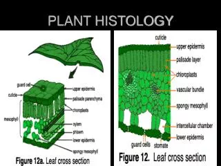
组织(tissue): 由细胞和细胞外基质 extracellular matrix构成。 人体(基本)组织:上皮组织、结缔组织、肌组织和神经组织---胚胎发生来源、细胞构成、形态特点和功能等方面,具有明显的特性。 • Tissue is composed of cells and extracellular matrix;collections of cells with similar morphological characteristics: • Original 4 types of (basic, primary, or fundamental) tissues: • Epithelial tissues – surface coverage • Muscular tissues – contractile property • Nervous tissues – cells forming brain, spinal cord, and nerves • Connective tissues – to link or support other specialized tissues • Tissues are different from each other in their origin, cellulosity细胞构成, morphological characteristics and functions.
器官organ:四种基本组织以不同的种类、数量和方式组合形成器官。器官organ:四种基本组织以不同的种类、数量和方式组合形成器官。 系统system:若干功能相关的器官构成系统。 Each of the fundamental tissues is formed by several types of cells and typically by specific associations of cells and extracellular matrix. These characteristic associations facilitate the recognition of the many subtypes of tissues. Most organs are formed by an orderly combination of several tissues, except the central nervous system, which is formed almost solely by nervous tissue. The precise combination of these tissues allows the functioning of each organ and of the organism as a whole. A system is a group of organs that work together and perform one or more functions.
细胞外基质extracellular matrix: The extracellular matrix (ECM) is the noncellularcomponent present within all tissues andorgans, and provides not only essential physicalscaffolding for the cellular constituents but alsoinitiates crucial biochemical and biomechanicalcues that are required for tissue morphogenesis,differentiation and homeostasis. The importanceof the ECM is vividly illustrated by the widerange of syndromes, which can be anythingfrom minor to severe, that arise from geneticabnormalities in ECM proteins. Although, fundamentally, theECM is composed of water, proteins andpolysaccharides, each tissue has an ECM witha unique composition and topology that isgenerated during tissue development througha dynamic and reciprocal, biochemicaland biophysical dialogue between thevarious cellular components (e.g. epithelial,fibroblast, adipocyte, endothelial elements)and the evolving cellular and proteinmicroenvironment.
细胞 Cell 构成:细胞膜、细胞质和细胞核,不同的细胞有不同的亚细胞结构特点。 The cell is the basic structural, functional and biological unit of all known living organisms. Cells are the smallest unit of life that is classified as a living thing, and are often called the "building blocks of life". Cells consist of a protoplasm enclosed within a membrane, the cell membrane or plasma membrane, which contains many biomolecules such as proteins and nucleic acids. Organisms can be classified as unicellular (consisting of a single cell; including most bacteria) or multicellular (including plants and animals). While the number of cells in plants and animals varies from species to species, humans contain about 100 trillion (1014) cells. Most plant and animal cells are between 1 and 100 micrometres and therefore are visible only under the microscope. The cytoplasm comprises cytosol (or intracellular fluid (ICF) or cytoplasmic matrix) – the gel-like substance enclosed within the cell membrane – and the organelles – the cell's internal sub-structures. Within the cells of eukaryote organisms the contents of the cell nucleus are separated from the cytoplasm, and are then called the nucleoplasm. The cytoplasm is about 70% to 90% water and usually colorless.
亚细胞结构: A subcellular structure is simply structures within a cell. They are smaller than a cell and commonly means within a cell. They are individual components of a cell that when put together forms a complete cell. Subcellular structures can only be seen by an electron microscope, either SEM or TEM microscopy. Examples of subcellular structures are organelles - Golgi apparatus, smooth and rough endoplasmic reticulum, nucleus and mitochondria. 各种分子,其中生物大分子,特别是核酸、蛋白质是决定细胞形态和功能的关键因素。 Molecules, among them the biomacromolecules, especially nucleic acid and proteins, determine forms and functions of cells.
胚胎学embryology: 研究个体发生、发育及其机制的学科,包括:胚胎早期发育和各器官系统发育,先天性畸形及其成因。 与组织学互相联系,但相互独立。 Embryology is a science which is about the development of an embryo from the fertilization of the ovum to the fetus stage, mainly concerning the developmental processes and mechanism of a human being, is divided into general embryology that studies the formations and development of germ cells, fertilization and early development of human embryo, and special embryology that studies the formation and malformations of different organs. It also studies the causes inducing malformations. Embryology and Histology are connected with each other, and are separated from each other. Conception5weeks
畸形学Teratology:研究致畸因子作用和先天性畸形的成因及预防的科学畸形学Teratology:研究致畸因子作用和先天性畸形的成因及预防的科学 Teratology is the study of abnormalities of physiological development. It is often thought of as the study of human birth defects, but it is much broader than that, taking in other non-birth developmental stages, including puberty; and other non-human life forms, including plants. A newer term developmental toxicity includes all manifestations of abnormal development, not only frank terata. These may include growth retardation or delayed mental development without any structural malformations. Anencephaly [ænən'sefəlɪ] Cyclopia
学习组织学与胚胎学的意义 • 学习其他相关学科的前提: • 病理学Histology and Pathology: • Histology: observe the conditions of cells and tissues under disease-free condition. • Pathology: observe the changes of cells and tissues by the disease processes. • 生理学 其他学科:儿科学、内科学、外科学、 妇产科学 科学研究: • Research • The subject no longer merely deals with the structure of the body; it also concerns itself with the body’s function. In fact, histology has a direct relationship to other disciplines and is essential for their understanding. • You will recognize the importance of this subject as they refer to the text later in your careers. An excellent example of this relationship will be evident when the reader learns about the histology of the kidney and realizes it is the intricate and almost sublime structure of that organ (down to the molecular level) that is responsible for the kidney’s ability to perform its function. Alterations of the kidney’s structure are responsible for a great number of life-threatening conditions.
This program includes lectures and laboratory practices. The fast developing Histology & Embryology is a main and fundamental course for different specialty of medicine, and makes the students know the basic knowledge and techniques, and acquire the ability to analyze and solve problems in further studies in other medical courses and clinic management, even in your future careers. The remainder of this chapter will discusses the methods used by histologists and embryologists.
二、当代组织学与胚胎学 Modern H & E 电子显微镜(electron microscope,简称电镜)的发明和超微结构的发现 • The German physicist Ernst Ruska and the electrical engineer Max Knoll constructed the prototype electron microscope in 1931, capable of four-hundred-power magnification; the apparatus was the first demonstration of the principles of electron microscopy. Two years later, in 1933, Ruska built an electron microscope that exceeded the resolution attainable with an optical (light) microscope. Moreover, Reinhold Rudenberg, the scientific director of Siemens-Schuckertwerke, obtained the patent for the electron microscope in May 1931.
分辨率达0.2nm。20年后发展了超薄切片术;扫描电镜出现。可以观察细胞膜、细胞器、染色体、细胞间纤维成分的超微结构(ultrastructure),发现了组织、器官中的大量新的细胞种类、各种细胞间的连接和空间配置关系---达到亚细胞水平。分辨率达0.2nm。20年后发展了超薄切片术;扫描电镜出现。可以观察细胞膜、细胞器、染色体、细胞间纤维成分的超微结构(ultrastructure),发现了组织、器官中的大量新的细胞种类、各种细胞间的连接和空间配置关系---达到亚细胞水平。 • The maximizing resolution power of an electron microscope may reach 0.2nm. Microtomy was developed, after that 20 years, and then, scanning electron microscope was invented. Subcellular structures, such as plasma membrane and organelles, were seenwith the aid of electron microscopy. The structures visible under the electron microscopeis termed ultrastructure. • 普通光学显微镜和电子显微镜观察常用长度单位: • 毫米 mm, millimeter;微米μm, micrometer;纳米nm, nanometer Length units commonly used in microscopy and EMare mm, μm (1/1000mm)and nm (1/1000 μ m).
近代组织学与胚胎学进展: • 改进、发展各种技术,大量使用新仪器、技术: • 图像分析仪image analyzer、共焦激光扫描显微镜Confocal Laser Scanning Microscope、流式细胞术flow cytometry, FCM,免疫组织化学Immunocytochemistry。 • With development and advance in sciences and the advent of new technology, many new instruments and technique were applied by histologists and embryologists. • Confocal Laser Scanning Microscope • Image analyzer • Flow cytometry, FCM • Immunocytochemistry, and so on
三、组织学与胚胎学的研究方法 Research methods used in Histology and Embryology: 组织学技术种类繁多,其原理涉及物理、化学、生物化学、免疫学、分子生物学等知识 The small size of cells and matrix components makes histology dependent on the use of microscopes. Advances in physics, chemistry, biochemistry, physiology, immunology, molecular biology and pathology—and the interactions among these fields—are essential for a better knowledge of tissue biology. Familiarity with the tools and methods of any branch of science is essential for a proper understanding of the subject. This chapter reviews some of the more common methods used to study cells and tissues and the principles involved in these methods.
(一)一般光学显微镜技术 Light (Optical) microscopy 常用光镜标本制备 石蜡切片(paraffin sectioning): ①取材和固定Tissue Specimen collection and fixation: 固定剂—蛋白质凝固剂:常用甲醛formaldehyde--在很大程度上保存组织的原本结构。组织块一般不超1.0cm大小。 • To avoid tissue digestion by enzymes present within the cells (autolysis) or by bacteria and to preserve the structure and molecular composition, pieces of organs should be promptly and adequately treated before or as soon as possible after removal from the body. This treatment—fixation —can be done by chemical or, less frequently, physical methods. In chemical fixation, the tissues are usually immersed in solutions of stabilizing or cross-linking agents called fixatives. Because the fixative needs some time to fully diffuse into the tissues, the tissues are usually cut into small fragments (about 1.0cm in length, width and thickness) before fixation to facilitate the penetration of the fixative and to guarantee preservation of the tissue. The most common fixative for light microscopy is 10% neutral bufferedformalin(4% formaldehydein phosphate buffered saline).
Tissue preparation A. paraffin section preparation • Specimen:as fresh as possible • Fixation:fixative: formalin solution; purpose: to preserve the structural organisation • Dehydration:replace the water in the tissue by alcohol • Clearing:replace the alcohol by xylene • Embedding: replace the xylene w/ melted paraffin • Sectioning & mounting l
The fixative preserves tissues or cells mainly by irreversibly cross-linking proteins. The main action of these aldehyde fixatives is to cross-link amino groups in proteins through the formation of methylene bridges (-CH2-), in the case of formaldehyde. This process, while preserving the structural integrity of the cells and tissue can damage the biological functionality of proteins, particularly enzymes, and can also denature them to a certain extent. This can be detrimental to certain histological techniques. Further fixatives are often used for electron microscopy such as osmium tetroxide or uranyl acetate. • Formalin fixation leads to degradation of mRNA, miRNA and DNA in tissues. However, extraction, amplification and analysis of these nucleic acids from formalin-fixed, paraffin-embedded tissues is possible using appropriate protocols.
②脱水dehydration和包埋embedding:梯度酒精ascending graded ethanol脱去固定好的组织块中的水分;二甲苯dimethylbenzene; xylene; xylol 置换组织中的酒精—不溶于石蜡;浸蜡—将组织块置于融化的石蜡paraffin; paraffin wax; paraffine中,让石蜡浸入组织细胞,冷却—蜡块—具有石蜡的硬度。 Biological specimens often need to be solidified to allow fine sectioning. Embedding materials include paraffin and plastic resins. Paraffin is used routinely for light microscopy; resins are used for both light and electron microscopy.
Tissue blocks are first stabilized by fixatives. Then: Dehydration: Blocks are dehydratedin ascending graded ethanol series (usually from 70% to 100% ethanol ). Clearing: The ethanol is then replaced with a solvent miscible with the embedding medium. In paraffin embedding, the solvent used is usually xylene. As the tissues are infiltrated with the solvent, they generally become transparent (clearing). Embedding: The blocks are embedded in paraffin wax which is the typical embedding medium for routine histology. • Once the tissue is impregnated with the solvent, it is placed in melted paraffin (paraffin wax) in the oven, typically at 58–60℃. The heat causes the solvent to evaporate, and the spaces within the tissues become filled with paraffin. The tissue together with its impregnating paraffin hardens after being taken out of the oven.
Tissue processing Embedding moulds: (A) paper boat; (B) metal boat mould; (C) Dimmock embedding mould; (D) Peel-a-way disposable mould; (E) base mould used with embedding ring ( F) or cassette bases (G)
③切片Sectioning和染色staining:用切片机将组织切为5-10μm薄片,贴在载玻片上,脱蜡,水化(由高到低浓度酒精descending grades of alcohol,到水),染色。 The hard blocks containing the tissues are then taken to a microtome and are sectioned by the microtome’s steel or glass blade to a thickness of 1-10μm. The sections are floated on water and transferred to glass slides to be stained. Because many tissue constituents have approximately the same optical densities, they must be stained for light microscopy, usually with watersoluble stains. Therefore, the paraffin must first be removed from the section, after which the tissue is rehydrated and stained.
After staining, the section isagain dehydrated so that the coverslip may be permanentlyaffixed by the use of a suitable mountingmedium. The coverslip not only protects the tissue fromdamage but also is necessary for viewing the sectionwith the microscope. • Various types of stains have been developed for visualizationof the many components of cells and tissues;they may be grouped into three classes: • Stains that differentiate between acidic and basiccomponents of the cell • Specialized stains that differentiate the fibrous componentsof the extracellular matrix • Metallic salts that precipitate on tissues, formingmetal deposits on them • The most commonly used stains in histology arehematoxylin and eosin (H&E).
④封片Mounting:切片经脱水—梯度酒精(由低浓度到高浓度),滴加树胶,盖玻片密封保存。④封片Mounting:切片经脱水—梯度酒精(由低浓度到高浓度),滴加树胶,盖玻片密封保存。 • The final step in this procedure is topermanently mount the sections under a coverslip. This is accomplished by covering the sectionin a medium that will harden and produce a clear binder between the slide and coverslip. Theideal mounting medium should not distort the stain color, or yellow and become brittle with age. • Rinsing, Dehydration & Mounting Prep • water • 70 % ethanol 2-3 minutes • 80 % ethanol 2-3 minutes • 95% ethanol2-3 minutes • 100% ethanol2-3 minutes • Clearing Agent 10 minute • Clearing Agent 10 minute • I used mounting resin called Permount (Fisher Scientific) and Neutral balsam.
How to get tissues for study? • Steps in tissue preparation • Fresh tissues from the body • 1. fixation Formalin ( 10% formaldehyde) • Osmium tetroxide for EM • Mechanism - Forms cross links with proteins (Lysine) • 2. Embedding – gives support for tissue slicing Paraffin or plastic resin • 3. Washing & dehydration (dehydration by graded alcohols in ascending order) • 4. clearing – to remove paraffin & alcohol By xylol • 5. block making
6. section cutting – 5-10μ thick sections with microtome • 7. mounting – on glass slide ( adhesive – albumin) • 8. clearing – xylol • 9. rehydrate – alcohols in descending order • staining nuclear stain – Hematoxylin ( basic stain & water soluble) • counter stain – Eosin ( less water soluble but soluble in alcohol) – dehydrate in ascending order • 10. dehydrate and Clearing • – alcohols in ascending order • – xylol • 11.Mounting medium – cover glass
Staining • Purpose: To make tissue section pigment for observation. • H-EStaining: Hematoxylin:basicdye,purple-blue Eosin: acid dye, pink color Basophilic: components bonded by basic dye (H); pruple-blue(nuclear chromatin & basophilic substance in cytoplasm) Acidophilic: components bonded by acidic dye (E); pink (cytoplasm & collagenous fiber)
Neutrophilic:do not stain w/ both basic and acid dyes Metachromasia:a dye stains tissue a different color from that of dye solution, e.g. toluidine blue stains mast cells in purple color
With few exceptions, most tissues are colorless, so observing them unstained in the light microscope is useless. Methods of staining tissues have therefore been devised that not only make the various tissue components conspicuous but also permit distinctions to be made between them. The dyes stain tissue components more or less selectively. Most of these dyes behave like acidic or basic compounds and have a tendency to form electrostatic (salt) linkages with ionizable radicals of the tissues. Tissue components, such as ribosomes that stain more readily with basic dyes are termed basophilic , so basophilia is the affinity of cellular structures for basic dyes, such as methylene blue; those with an affinity for acid dyes are termed acidophilic, acidophilia is the affinity of cellular structures for acidic dyes, such as eosin. Those with an weak affinity for both basic dyes and acid dyes are termed neutrophilic, neutrophilia is the affinity of cellular structures for neutral dyes • Hematoxylin behaves like a basic dye, that is, it stains the basophilic tissue components. The main tissue components that ionize and react with basic dyes do so because of acids in their composition (nucleic acids and acid glycoproteins). Acid dyes (eg, orange G, eosin, acid fuchsin) stain the acidophilic components of tissues such as mitochondria, secretory granules, and collagen.
苏木精染色液 二甲苯1 二甲苯2 100%酒精 100%酒精 95%酒精 95%酒精 85%酒精 75%酒精 水洗 盐酸酒精 在染色缸中 水洗 二甲苯2 二甲苯1 100%酒精 100%酒精 95%酒精 95%酒精 85%酒精 75%酒精 伊红染色液 • 苏木精—伊红染色法(HE染色法) (hematoxylin-eosin staining)
Hematoxylin and Eosin (H & E) Staining • H & E is a charge-based, general purpose stain. Hematoxylin stains acidic molecules shades of blue. Eosin stains basic materials shades of red, pink and orange. H & E stains are universally used for routine histological examination of tissue sections. • Fixation • Any well fixed tissue. • Staining Procedure • 1- Deparaffinize and hydrate to water • 2- If sections are Zenker-fixed, remove the mercuric chloride crystals with iodine and clear with sodium thiosulphate (hypo)
3- Hematoxylin for 15 minutes • 4- Wash in running tap water for 20 minutes • 5- Counterstain with eosin from 15 seconds to 2 minutes depending on the age of the eosin, and the depth of the counterstain desired. For even staining results dip slides several times before allowing them to set in the eosin for the desired time • 6- Dehydrate in 95% and absolute alcohols, two changes of 2 minutes each or until excess eosin is removed. Check under microscope • 7- Clear in xylene, two changes of 2 minutes each • 8- Mount in Permount or Histoclad • Results • Nuclei - blue - with some metachromasia • Cytoplasm - various shades of pink-identifying different tissue components
切片及染色设备 • 自动染色机 切片机及其它设备 Microtome for sectioning resin- and paraffin-embedded tissues for light microscopy. Rotation of the drive wheel moves the tissue-block holder up and down. Each turn of the drive wheel advances the specimen holder a controlled distance, generally between 1 and 10μ m. After each forward move, the tissue block passes over the knife edge, which cuts the sections.
其他: • 火棉胶包埋celloidin embedding,火棉胶切片机切片:制作大组织块切片。 • Nitrocellulose, otherwise known as cellulose nitrate, goes by several names. The commonest, in the histological world, are celloidin and collodion. Tissues embedded in celloidin are usually sectioned with a sliding microtome. In this instrument the block is mounted on a platform facing upwards and is fixed. The knife is held at a significant slant so that most of the blade edge is used during the cutting stroke, and is quite long, often in excess of 25 cm.
Cryostat are used in medicine to cut histological slides. They are usually used in a process called frozen section histology. The cryostat is essentially an ultrafine "deli-slicer", called a microtome, placed in a freezer. The cryostat is usually a stationary upright freezer, with an external wheel for rotating the microtome. The temperature can be varied, depending on the tissue being cut - usually from minus 20 to minus 30 degree Celsius. The freezer is either powered by electricity, or by a refrigerant like liquid nitrogen. • 冰冻切片frozen section: • 新鲜组织,液氮liquid nitrogen(-1960C)冷冻,恒冷箱切片机cryostat microtome—检查组织化学、酶等
1. Freeze a fresh, unfixed tissue sample, up to 2.0 cm in diameter, in OCT in a suitable tissue mold. Freeze the OCT containing the tissue onto the specialized metal grids that fit onto the cryostat. • OCT is viscous at room temperature and miscible with H2O, but freezes into a solid support at −20°C. • Certain soft tissues, such as brain, are optimally frozen in M-1 medium at −3°C. • 2. Cut sections 5-15 μm thick in the cryostat at −20°C. If necessary, adjust the temperature of the cutting chamber ±5°C, according to the tissue under study. • OCT :Optimal Cutting Temperature Compound, the formulation of water-soluble glycols and resins, is used to embed tissue samples prior to frozen sectioning on a microtome-cryostat. This process is undertaken so as to mount slices (sections) of a sample onto slides for analysis.
特殊染色Histology Special Stains • 硝酸银(镀银)染色Silver impregnation, silver staining,染神经细胞,纤维成分等。 某些结构经硝酸银处理使硝酸银还原,形成银微粒附在组织结构上-棕黑色,此性质称亲银性(argentaffin)。有些组织结构不能使硝酸银还原,需加还原剂使硝酸银还原,称为嗜银性(argyrophilia)。 Silver (Silver nitrate)staining( Silver impregnation ) aids the visualisation of targets of interest, namely intracellular and extracellular cellular components such as DNA and proteins, such as type III collagen and reticulin fibres by the deposition of metallic silver particles on the targets of interest.
Argentaffin:Of or relating to cells or tissue elements that reduce silver ions in solution, thereby staining brown or black. In the argentaffin reaction, the tissue contains reducing groups that are sufficiently strong and present in sufficient quantity to give a visible depositwithout added reducing agents.These groups are often aldehyde groups and silver solutions can be used to replace the Schiff ’s reagent in the PAS technique to give periodic acid–silver. Argyrophilia: Many tissue groups are able to adsorb silver, possibly by ionic mechanisms as for dyeing. The silver is mainly adsorbed as silver ions but small amounts are reduced to silver atoms. These silver atoms are deposited at the site of reduction. The initial reduction reaction with silver only deposits submicroscopic atoms of silver at particularly reactive sites. In this case the developer does the main reduction and the tissue simply provides places where there are silver atoms to catalyse the reduction. This type of reaction where an external reducer or developer is added is called an argyrophilic reaction.
异染性metachromasia: 用甲苯胺蓝等碱性染料染色呈紫红色。 • The term metachromasia is used when a dye stains a tissue component a different color to the dye solution. For example, toluidine blue is a strong basic blue dye that stains nuclei a deep blue color; however, it will also stain mast cell granules a pink color. This color shift that occurs with mast cells is called metachromasia, whilst the usual blue staining is called orthochromasia.Many dyes can show metachromasia but the thiazine group dyes are especially good for this type of staining.
Silver staining of the neuron and the bile canaliculi HO (Hoechst 33258)-PI staining shows the apoptosis in human HL-60 cells
涂片smear:直接将细胞(如血细胞)涂于玻片上,制作血涂片、细胞涂片等。涂片smear:直接将细胞(如血细胞)涂于玻片上,制作血涂片、细胞涂片等。 Smear:A small quantity of cells (e.g. blood cells, cells collected by a swab or cultured) spread thinly on a slide for microscopic examination. • 铺片:将组织撕成薄片铺在载玻片上,制作结缔组织铺片、肠系膜铺片等。 Whole-mounts, stretched preparation, where an entire organism or structure is small enough or thin enough to be placed directly onto a microscope slide (e.g. a small unicellular or multicellular organism or a membrane that can be stretched thinly on to a slide)
磨片ground section:将硬组织磨成薄片,制作骨磨片、牙磨片等。 A ground section, (e.g. bone), refers to a microscope slide of bone that is prepared by taking a larger piece of the bone and placing it between two pieces of abrasive material--such as carbide paper. These are rotated and "grind" the section down until the bone is adequately thin to transmit incident light in a light microscope, allowing for observation of the bone structure. This section is then transferred to a glass slide, mounted, and cover slipped.
显微镜: Microscopy Light microscopy Conventional light, phase contrast, differential interference, polarizing, confocal, and fluorescence microscopy are all based on the interaction of light and tissue components. With the light microscope, stained preparations are usually examined by means of light that passes through the specimen. The microscope is composed of mechanical and optical parts. The optical components consist of 3 systems: condenser, objective, and eyepiece. The condenser collects and focuses light, producing a cone of light that illuminates the object to be observed. The objective lenses enlarge and project the illuminated image of the object in the direction of the microscope. The microscope further magnifies this image and projects it onto the viewer’s retina, a photographic plate, or ( to obtained by digital image) a detector such as a charged coupled device (CCD电荷耦合设备) camera. The total magnification is obtained by multiplying the magnifying power of the objective and ocular lenses.
3、荧光显微镜(fluorescence microscope): 使用荧光染料染色或作为标记物,以紫外线为光源,激发染料发出荧光。