Download
1 / 40
420 likes | 771 Vues
SALIVARY GLAND DISEASES. Yrd . Doç.Dr . Rasim Yılmazer. Learning goal and objectives of the lesson. Learning goal of the lesson: The learner should know the main clinical features and investigation of salivary gland disorders Learning objectives of the lesson the learner will be able to:

E N D
SALIVARY GLAND DISEASES Yrd.Doç.Dr. Rasim Yılmazer
Learning goal and objectives of the lesson Learning goal of the lesson: The learner should know the main clinical features and investigation of salivary gland disorders Learning objectives of the lesson the learner will be able to: • identify the most common etiologies of salivary masses based on history and physical exam • develop a clear concinse algorithm for use of diagnostic tests evaluation of salivary masses • describe available therapeutic options for malignant salivary gland masses • Understand how to approach the patient with “ a lump in the parotis or submandibular gland. Skill objectives of the lesson the learner will be able to take a directed history and perform a physical exam on a patient with salivary gland masses.
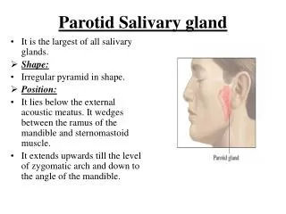
Introduction • Salivarygland is anycellor organ discharging a secretioninto oral cavity • Major (paired) • Parotid (Stensen’sduct) • Submandibular (wharton’sduct • Sublingual • Minor • Those in tongue, palatinetonsil, palate, lipsandcheeks
Mumps • Most common viral disorder of salivary glands • Peak age 4-6 • Prodrome period is 2-3 weeks • 1 or both parotid glandes can be involved • Contagious from approximately 6 days before the onset of symptoms until about 9 days after symptoms start • Diagnosis: diagnosed on clinical grounds • Serum amylase is often elevated
Complications: deafness, pancreatitis, meningitis, encephalitis,orchitis or epididymitis, Oophoritis (inflammation of ovaries) • Deafness generally unilateral rarely bilateral • Profound (91 dB or more) sensorineural hearing loss • Acute unilateral deafness occurs in about 0.005% of cases
No specific treatment • Paracetamol for pain relief • Warm saltwater gargles, soft foods, and extra fluids may also help relieve symptoms • Self-limiting, and general outcome is good • Most common preventative measure against mumps is a vaccination with a mumps vaccine
Other Viruses • CMV, Coxsackievirus A, Echovirus, Influenza A, Lymphocytic choriomeningitis Virus • Treatment: symptomatic for all viral diseases
Acute bacterial parotitis • most often caused by a bacterial infection of Staphylococcus aureus but may be caused by any commensal bacteria • Peak age 50’s-60’s • 30-40% in post-op patients; most commonly gastrointestinal procedures
Presentation: sudden, diffuse enlargement with associated induration and tenderness. Massage produces purulent saliva • 20% of cases bilateral • Treatment: hydration, improved oral hygiene, repeated massage of gland, IV antibiotics, warm compresses, sialogogues • If no significant improvement in 24-48h, then proceed to incision & drainage OR image-guided needle aspiration
Chronic NonspecificSialadenitis • Most commonly parotid • Usually from permanent damage during acute infection; occasionally from recurrent parotitis of childhood
Recurrent Parotitis of Childhood • More common in males; peak age 5-7 • ¾ give role of Mumps; heredity plays no role • Presentation: Usually unilateral; when bilateral, one side worse Severe pain, fever, malaise during attacks Recurs 55% of cases resolve with puberty 25% no improvement with puberty
Sarcoidosis • Parotid enlargement is a classic feature of sarcoidosis, but clinically apparent parotid involvement occurs in less than 10% of patients. • Bilateral involvement is the rule. • The gland is usually not tender, but firm and smooth. • Xerostomia can occur • Other exocrine glands are affected only rarely
Sjogren’s Syndrome: • Sjögren syndrome also known as "Sicca syndrome" • Systemic autoimmune disease in which immune cells attack and destroy the exocrine glands that produce tears and saliva • Chronic, slowly progressive, benign; 2nd most common autoimmune disease behind Rheumatoid arthritis
AlthoughSjögren's occurs in all age groups in both women and men • Nine out of ten Sjögren's patients are women • Average age of onset is after menopause in women
Presentation • Other exocrine gland involvement: dry nose, dry throat, xerotrachea, esophageal mucosal atrophy, atrophic gastritis, subclinical pancreatitis, vaginal dryness • 1/3 = fatigue, low grade fever, myalgias/arthralgias • Extraglandular involvement in ¼: Lungs, kidneys, vasculitis, nervous system
Associated risks • Increased risk of 1) NonHodgkin’s Lymphoma 2) Multiple Myeloma
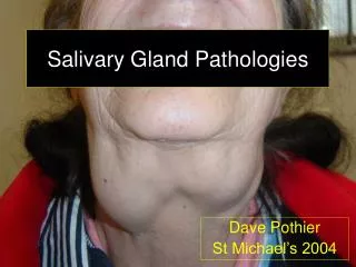
Sialolithiasis • Formation of stones in the salivary glands • 80% submandibular gland, 20% parotid • Only 1 stone in ¾ cases • Presentation: recurrent swelling, pain worse with eating • Complications: sialadenitis, ductalectasia, and stricture
Diagnosis is usually made by characteristic history and physical examination • Diagnosis can be confirmed by x-ray (80% of salivary gland calculi are visible on x-ray), or by sialogram or ultrasound. • 90% of submandibular stones radioopaque; 90% of parotid stones radiolucent • Treatment: Endoscopicexcision, If near duct orifice transoral removal of stone with marsupialization, gland excision
Cysts • Mucous cysts: minor salivary glands • Mucocel2-5% of all parotid lesions • Congenital: dermoid cysts, ductal cysts, 1st arch branchial cleft cysts • Acquired: trauma, parotitis, calculi, neoplasms
Benign Neoplasms Pleomorphic Adenoma Warthin’s Tumor Oncocytoma Monomorphic Adenomas Myoepithelioma Malignant Neoplasms Mucoepidermoid Carcinoma Adenoid Cystic Carcinoma Acinic Cell Carcinoma Adenocarcinoma Malignant Mixed Tumor Squamous Cell Carcinoma Clear Cell Carcinoma Epithelial-Myoepithelial Carcinoma Salivary Gland Neoplasms
Salivary Gland Neoplasms • Diverse histopathology • Relatively uncommon • 2% of head and neck neoplasms • Distribution • Parotid: 80% overall; 80% benign • Submandibular: 15% overall; 50% benign • Sublingual/Minor: 5% overall; 40% benign
Pleomorphic Adenoma • Most common of all salivary gland neoplasms • 70% of parotid tumors • 50% of submandibular tumors • 45% of minor salivary gland tumors • 6% of sublingual tumors • 4th-6th decades • F:M = 3-4:1
Pleomorphic Adenoma • Slow-growing, painless mass • Parotid: 90% in superficial lobe, most in tail of gland • Minor salivary gland: lateral palate, submucosal mass
Pleomorphic Adenoma • Treatment: complete surgical excision • Parotidectomy with facial nerve preservation • Submandibular gland excision • Wide local excision of minor salivary gland • Avoid enucleation and tumor spill
Pleomorphic Adenoma • Gross pathology • Smooth • Well-demarcated • Solid • Cystic changes • Myxoid stroma
Warthin’s Tumor • Papillarycystadenomalymphomatosum • 6-10% of parotid neoplasms • Older, caucasian, males • 10% bilateral or multicentric • 3% with associated neoplasms • Presentation: slow-growing, painless mass
Oncocytoma • Rare: 2.3% of benign salivary tumors • 6th decade • M:F = 1:1 • Parotid: 78% • Submandibular gland: 9% • Minor salivary glands: palate, buccal mucosa, tongue
Monomorphic Adenomas • Basal cell, canalicular, sebaceous, glycogen-rich, clear cell • Basal cell is most common: 1.8% of benign epithelial salivary gland neoplasms • 6th decade • M:F = approximately 1:1 • Caucasian > African American • Most common in parotid
Mucoepidermoid Carcinoma • Most common salivary gland malignancy • 5-9% of salivary neoplasms • Parotid 45-70% of cases • Palate 18% • 3rd-8th decades, peak in 5th decade • F>M • Caucasian > African American
Mucoepidermoid Carcinoma • Presentation • Low-grade: slow growing, painless mass • High-grade: rapidly enlarging, +/- pain
Mucoepidermoid Carcinoma • Gross pathology • Well-circumscribed to partially encapsulated to unencapsulated • Solid tumor with cystic spaces
Adenoid Cystic Carcinoma • Overall 2nd most common malignancy • Most common in submandibular, sublingual and minor salivary glands • M = F • 5th decade • Presentation • Asymptomatic enlarging mass • Pain, paresthesias, facial weakness/paralysis
Acinic Cell Carcinoma • 2nd most common parotid and pediatric malignancy • 5th decade • F>M • Bilateral parotid disease in 3% • Presentation • Solitary, slow-growing, often painless mass
Adenocarcinoma • Rare • 5th to 8th decades • F > M • Parotid and minor salivary glands • Presentation: • Enlarging mass • 25% with pain or facial weakness
Malignant Mixed Tumors • Carcinoma ex-pleomorphic adenoma • Carcinoma developing in the epithelial component of preexisting pleomorphic adenoma • Carcinosarcoma • True malignant mixed tumor—carcinomatous and sarcomatous components • Metastatic mixed tumor • Metastatic deposits of otherwise typical pleomorphic adenoma
Squamous Cell Carcinoma • Gross pathology • Unencapsulated • Ulcerated • fixed