Download
1 / 68
700 likes | 901 Vues
Update in Acute Stroke: Treatment and Prevention. AOA/OMED 10/1/13 Eric P. Baron, DO Cleveland Clinic Neurological Institute Center for Regional Neurology. Stroke FACTS.

E N D
Update in Acute Stroke: Treatment and Prevention AOA/OMED 10/1/13 Eric P. Baron, DO Cleveland Clinic Neurological Institute Center for Regional Neurology
Stroke FACTS • In 2008, fell from 3rd to 4th leading cause of death in the US after heart disease, cancer, and chronic lower respiratory diseases • >795,000 people in US have a stroke each year. -About 610,000 of these are first or new strokes. -1 in 4 are recurrent strokes. • >137,000 deaths each year (1 in every 18 deaths) http://www.strokeassociation.org/STROKEORG/AboutStroke/Impact-of-Stroke-Stroke-statistics_UCM_310728_Article.jsp
STROKE FACTS • About 40% of stroke deaths occur in males, and 60% in females. • An American has a stroke every 40 seconds, and every 4 minutes someone dies of stroke. • Stroke costs the US >$38.6 billion per year http://www.strokeassociation.org/STROKEORG/AboutStroke/Impact-of-Stroke-Stroke-statistics_UCM_310728_Article.jsp
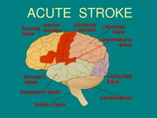
Types of Stroke Ischemic Stroke (87%) A thrombus or embolus blocks blood flow to part of the brain. Hemorrhagic Stroke (13%) Blood spills out from break in blood vessel in brain
Stroke Types • Ischemic = 87% - 45% Atherothrombotic - 20% Cardioembolic - 20% Cryptogenic - 2% Other • Hemorrhagic = 13% - 10% Intracerebral hemorrhage - 3% Subarachnoid hemorrhage
Cerebral Ischemia • Each hour: -120 million neurons, 830 billion synapses, and 714 km (447 miles) of myelinated fibers are lost. -Compared with normal rate of neuron loss in brain aging, the ischemic brain ages 3.6 years each hour without treatment. • Each minute: -1.9 million neurons, 14 billion synapses, and 12 km (7.5 miles) of myelinated fibers are destroyed. • Each second: -30,000 neurons
Transient Ischemic Attack (TIA) • Classical definition (1960s): Sudden, neurological deficit, usually focal, lasting < 24 hours. NIH Consensus Statement. Stroke 1975;6:564-616. • Newer definition (2002): Sudden neurological deficit lasting less than 1 hour and not associated with new lesion on neuroimaging. (e.g. DWI on MRI) Albers GW et al. N Eng J Med 2002;347(21):1713-1716. • New AHA endorsed definition (2009): A transient episode of neurological dysfunction caused by focal brain, spinal cord, or retinal ischemia, without acute infarction. Easton JD et al. Stroke 2009;40:2276-2293.
Transient Ischemic Attack (TIA) • Reversible neurologic deficits typically <30 minutes and usually resolve within 1 hour, although may last up to 24 hours. • Often called a “mini-stroke” • Due to a temporary disruption of the blood supply to the brain • WARNING SIGN FOR STROKE
Transient Ischemic Attack (TIA) • Risk of stroke after TIA - 1/3 have continued TIAs - 1/3 have no further symptoms - 1/3 stroke within 5 years (usually 1st month) - 50% have stroke in next 2 days - 10% have stroke in next 3 months - 15% have stroke in 1 year - 25% have stroke in 5 years
http://www.theanswerpage.com/qod.php?specialty_id=4&day=06/26/2012http://www.theanswerpage.com/qod.php?specialty_id=4&day=06/26/2012
Transient Ischemic Attack (TIA) • 40-60% of TIA ptshave evidence of ischemic injury on DWI (Diffusion Weighted Imaging) • Factors predicting positive DWI: • Symptoms lasting > 60 minutes • Focal weakness • Speech impairment Kidwell C et al. Stroke 1999; 6:1174-1180. Couttts SB et al. Annals of Neurology 2005;57:848-854
Class I Recommendations Patients with TIA should preferably undergo neuroimaging evaluation within 24 hours of symptom onset. MRI, including DWI, is the preferred brain diagnostic imaging modality. If MRI is not available, head CT should be performed (Class I, LOE B). Noninvasive imaging of the cervicocephalic vessels should be performed routinely as part of the evaluation of patients with suspected TIAs (Class I, LOE A). Easton JD et al. Stroke. 2009;40:2276-2293.
Class II Recommendations Initial assessment of the extracranial vasculature may involve any of the following: carotid ultrasound/TCD, MRA or CTA, depending on local availability and expertise, and characteristics of the patient (Class IIa, Level of Evidence B). If only noninvasive testing is performed prior to endarterectomy, it is reasonable to pursue two concordant noninvasive findings; otherwise catheter angiography should be considered (Class IIa, Level of Evidence B). Easton JD et al. Stroke. 2009;40:2276-2293.
Class II Recommendations Electrocardiography should occur as soon as possible after TIA (Class I, Level of Evidence B). Prolonged cardiac monitoring (inpatient telemetry or Holter monitor) is useful in patients with an unclear etiology after initial brain imaging and electrocardiography (Class IIa, LOE B). Echocardiography (at least TTE) is reasonable in the evaluation of patients with suspected TIAs, especially when the patient has no cause is identified by other elements of the work-up (Class IIa, Level of Evidence B). TEE is useful in identifying patent foramen ovale, aortic arch atherosclerosis, and valvular disease and is reasonable when identification of these conditions will alter management (Class IIa, LOE B). Easton JD et al. Stroke. 2009;40:2276-2293.
Class II Recommendations It is reasonable to hospitalize patients with TIA if they present within 72 hours of the event and any of the following criteria are present: ABCD2 score of ≥3, (Class IIa, LOE C). ABCD2 score of 0-2 and uncertainty that diagnostic work-up can be completed within 2 days as an outpatient (Class IIa, LOE C). ABCD2 score of 0-2 and there is other evidence that indicates the patient’s event was caused by focal ischemia (Class IIa, LOE C). Easton JD et al. Stroke. 2009;40:2276-2293.
Modifiable Risk Factors • Hypertension (high blood pressure) • Exposure to cigarette smoking (active or passive) • Diabetes • Atrial fibrillation and other cardiac conditions • Hyperlipidemia (high cholesterol) • Carotid artery stenosis • Sickle cell disease • Postmenopausal hormone therapy • Poor Diet • Physical Inactivity • Obesity and body fat distribution
Hypertension • Most important risk factor for stroke. • Causes a 2-4 fold increase in the risk of stroke before age 80.
Cholesterol Recommendations in Stroke • Prior Stroke or TIA: - LDL < 100 • Known coronary heart disease or coronary heart disease risk equivalents (diabetes, peripheral arterial disease, abdominal aortic aneurysm, or carotid artery disease): - LDL < 70
Smoking • Causes a 2-fold increase in risk of ischemic stroke and up to a 4-fold increase in risk of hemorrhagic stroke. • Linked to the buildup of atherosclerosis. • Nicotine raises BP; CO from smoking reduces the amount of oxygen blood can carry to the brain; and cigarette smoke makes blood thicker and more likely to clot. • Promotes aneurysm formation. • Stopping smoking causes a 50% risk reduction for stroke within 1 year. • Stopping smoking causes stroke risk to approach that of nonsmokers after 5 years.
DIABETES • Having diabetes is the equivalent of aging 15 years. • Causes destructive changes in the blood vessels throughout the body, including the brain. • If blood glucose levels are high at the time of a stroke, then brain damage is usually more severe and extensive than when blood glucose is well-controlled. • HTN is common among diabetics and accounts for much of the increased stroke risk.
Atrial fibrillation • In general, increases stroke risk by 5-fold. • Responsible for 1 in 4 strokes after age 80, and is associated with higher mortality and disability. • CHADS2 Score is helpful in deciding on anticoagulation: -Low risk (0): Aspirin 100-300 mg daily -Moderate risk (1): Warfarin or Aspirin -High risk (2-6): Warfarin (INR 2-3)
Prevention of 1st and Recurrent Stroke in Nonvalvular A-fib (AHA/ASA 8/2012 Recommendations) • Warfarin (Class I; Level of Evidence A) • Dabigatran 150 mg bid (Class I; Level of Evidence B) -CrCl >30 mL/min • Apixaban 5 mg bid (Class I; Level of Evidence B) -No more than 1 of the following characteristics: Age >80 years, weight <60 kg, creatinine >1.5 mg/dL • Rivaroxaban 20 mg/d (Class IIa; Level of Evidence B) -CrCl >50
Symptomatic extracranial carotid disease • Recent TIA or ischemic stroke w/in 6 months and ipsilateral severe (70%-99%) carotid stenosis: -CEA recommended if perioperative morbidity and mortality risk estimated at <6% (Class I; Level of Evidence A). • Recent TIA or ischemic stroke and ipsilateral moderate (50%-69%) carotid stenosis: -CEA recommended depending on patient-specific factors such as age, sex, and comorbidities if perioperative morbidity and mortality risk estimated at <6% (Class I; Level of Evidence B).
Symptomatic extracranial carotid disease • Stenosis <50%: -No indication for carotid revascularization by either CEA or CAS (Class III; Level of Evidence A). • When CEA is indicated for patients with TIA or stroke, surgery w/in 2 weeks is reasonable rather than delaying surgery if no contraindications to early revascularization (Class IIa; Level of Evidence B).
Symptomatic extracranial carotid disease • CAS indicated as alternative to CEA for symptomatic patients at average to low risk of complications associated with endovascular intervention when ICA stenosis >70% by noninvasive imaging or >50% by catheter angiography (Class I; Level of Evidence B).
Symptomatic extracranial carotid disease • Among patients with symptomatic severe stenosis (>70%) in whom the stenosis is difficult to access surgically, medical conditions are present that greatly increase the risk for surgery, or when other specific circumstances exist, such as radiation-induced stenosis or restenosis after CEA, CAS may be considered (Class IIb; Level of Evidence B). • CAS in the above setting is reasonable when performed by operators with established periprocedural morbidity and mortality rates of 4-6%, similar to those observed in trials of CEA and CAS (Class IIa; Level of Evidence B).
Symptomatic extracranial carotid disease • For patients with symptomatic extracranial carotid occlusion, EC/IC bypass surgery is not routinely recommended (Class III; Level of Evidence A). • Optimal medical therapy, which should include antiplatelet therapy, statin therapy, and risk factor modification, is recommended for all patients with carotid artery stenosis and a TIA or stroke as outlined elsewhere in this guideline (Class I; Level of Evidence B).
Extracranial Vertebrobasilar Disease • Optimal medical therapy, which should include antiplatelet therapy, statin therapy, and risk factor modification, is recommended for all patients with vertebral artery stenosis and a TIA or stroke as outlined elsewhere in this guideline (Class I; Level of Evidence B). • Endovascular and surgical treatment of patients with extracranial vertebral stenosis may be considered when patients are having symptoms despite optimal medical treatment (including antithrombotics, statins, and relevant risk factor control) (Class IIb; Level of Evidence C)
Intracranial Atherosclerosis • For patients with a stroke or TIA due to 50%-99% stenosis of a major intracranial artery, aspirin is recommended in preference to warfarin (Class I; Level of Evidence B). - Patients in the WASID trial were treated with aspirin 1300 mg/d, but the optimal dose of aspirin in this population has not been determined. - On the basis of data on general safety and efficacy, aspirin doses of 50 mg/d to 325 mg/d are recommended (Class I; Level of Evidence B).
Intracranial Atherosclerosis • For patients with stroke or TIA due to 50% to 99% stenosis of a major intracranial artery, long-term maintenance of BP <140/90 mm Hg and total cholesterol level <200 mg/dL may be reasonable (Class IIb; Level of Evidence B).
Intracranial Atherosclerosis • For patients with stroke or TIA due to 50%-99% stenosis of a major intracranial artery, the usefulness of angioplasty and/or stent placement is unknown and is considered investigational (Class IIb; Level of Evidence C). • For patients with stroke or TIA due to 50%-99% stenosis of a major intracranial artery, EC/IC bypass surgery is not recommended (Class III; Level of Evidence B).
NoN-MODIFIABLE RISK FACTORS • Age • Sex • Low birth weight • Race/ethnicity • Genetic predisposition
Age • Stroke occurs in all age groups, including childhood or adolescence. • Studies show the risk of stroke doubles for each decade between the ages of 55 and 85.
Gender • Men have a higher risk for stroke, but more women die from stroke. • Men generally do not live as long as women, so men are usually younger when they have their strokes and therefore have a higher rate of survival.
Race • In African Americans stroke is more common and more deadly across all age groups. • Studies show that the age-adjusted incidence of stroke is about twice as high in African Americans and Hispanic Americans as in Caucasians. • An important risk factor for African-Americans is sickle cell disease, which can cause a narrowing of arteries and disrupt blood flow.
Family History • Members of a family might have a genetic tendency for stroke risk factors, such as an inherited predisposition for HTN, HLP, DM, etc. • The influence of a common lifestyle among family members also could contribute to familial stroke.
Stroke and TIA Treatment • Stroke risk factor modifications (including antiplatelet therapy (or anticoagulation if indicated), HLP, HTN, DM, Smoking, etc.): -Primary (prior to an event) -Secondary (following an event) • Improving patient education for rapid symptom evaluation due to short intervention treatment windows (tPA)
Signs of Stroke Severe headache with no known cause Trouble seeing in one or both eyes Confusion, trouble speaking or understanding Numbness or weakness of face, arm or leg, especially on one side Trouble walking, dizziness, loss of balance or coordination
Acute Stroke Treatment:General Supportive Measures • Cardiac monitoring • BP - <180/105 following tPA and lower BP by 15% per day - <220/120 if no tPA and lower BP by 15% per day • Airway support as needed and O2 sats >94% • Temp < 38°C (100.4°F) • Treat hypoglycemia (<60 mg/dL) and hyperglycemia (optimal range 140-180 mg/dL) • Treat hypovolemia with IV NS • Head of bed 15-30°
tPA (Tissue Plasminogen Activator) • Catalyzes the conversion of plasminogen to plasmin, the major enzyme responsible for clot breakdown. • 0.9 mg/kg IV (max dose 90 mg) -10% given as a bolus over 1 minute -Remaining 90% continuous infusion over 60 minutes • No antiplatelet/anticoagulants x 24 hours post-tPA