DNA
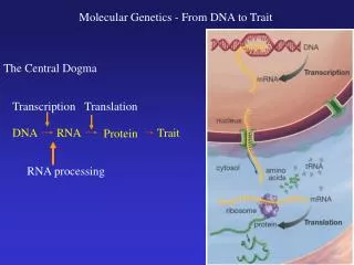
DNA. DNA. How DNA was discovered. Scientists that determined Structure and Importance of DNA. 1866 Gregor Mendel – offspring receive traits carried in a molecule 1869 Friedrich Meisher – isolated acid from cell nucleus – named it nucleic acid

DNA
E N D
Presentation Transcript
DNA DNA
Scientists that determined Structure and Importance of DNA 1866Gregor Mendel – offspring receive traits carried in a molecule 1869 Friedrich Meisher – isolated acid from cell nucleus – named it nucleic acid 1889 R.A. Altman-determined the chemical composition of the nucleic acid, DNA
Scientists that determined Structure and Importance of DNA 1919Phoebus Levene – Determined the structure of a unit of DNA called a nucleotide, including the sugar composition of DNA – first to propose that DNA was a polymer made of nucleotides P= Phosphate S= 5C sugar B= Nitrogen base
1928 -Frederick Griffith Took one of the most important steps toward finding DNA by trying to find better ways to fight pneumonia. Working withbacteriathat caused pneumonia, he discovered transformation
Transformation Process by which onestrain of bacteria is changed by a gene or genes from another strain of bacteria
Griffith isolated two different strains of the same bacterial species, but only one of the strains caused pneumonia. The disease-causing bacteria (S strain) grew into smooth colonies on culture plates, whereas the harmless bacteria (R strain) produced colonies with rough edges.
When Griffith injected mice with disease-causing bacteria, the mice developed pneumonia and died. When he injected mice with harmless bacteria, the mice stayed healthy. Perhaps the S-strain bacteria produced a toxin that made the mice sick? To find out, Griffith ran a series of experiments.
First, Griffith took a culture of the S strain, heated the cells to kill them, and then injected the heat-killed bacteria into laboratory mice. The mice survived, suggesting that the cause of pneumonia was not a toxin produced by these disease-causing bacteria.
In Griffith’s next experiment, he mixed the heat-killed, S-strain bacteria with live, harmless bacteria from the R strain and injected the mixture into laboratory mice. The injected mice developed pneumonia, and many died.
The lungs of these mice were filled with the disease-causing bacteria. How could that happen if the S strain cells were dead?
Griffith reasoned that some chemical factor that could change harmless bacteria into disease-causing bacteria was transferred from the heat-killed cells of the S strain into the live cells of the R strain. He called this process transformation, because one type of bacteria had been changed permanently into another.
Because the ability to cause disease was inherited by the offspring of the transformed bacteria, Griffith concluded that the transforming factor had to be a gene. R
1944-Oswald Avery A group of scientists at the Rockefeller Institute in New York, led by the Canadian biologist Oswald Avery, wanted to determine which molecule in the heat-killed bacteria was most important for transformation.
Avery and his team extracted a mixture of various molecules from the heat-killed bacteria and treated this mixture with enzymes that destroyed proteins, lipids, carbohydrates, and some other molecules, including the nucleic acid RNA. Transformation still occurred.
When DNA was destroyed, he discovered that transformation did not occur Conclusion: Genes are made from DNA
1949-Erwin Chargaff When studying the chemistry of DNA, he noticed each sample of DNA always contained equalamounts of adenine (A) and thymine (T), and equal amountsof cytosine (C) and guanine (G)
Rules: Adenine binds to Thymine (A to T) and Cytosine bind to Guanine (C to G)
Hershey – Chase Experiment In 1952, the work of two scientists, Hershey and Chase, with bacteriophages confirmed Avery’s results, convincing many scientists that DNA was the genetic material found in genes—not just in viruses and bacteria, but in all living cells.
Hershey and Chase studied viruses—nonliving particlesthat can infectlivingcells.
The kind of virus that infects bacteria is known as a bacteriophage,which means “bacteria eater.”
Bacteriophages • 1. attach to the surface of the bacterial cell and injects its genetic information into it. • 2. genes act to produce many new bacteriophages, which gradually destroy the bacterium. • 3. the cell splits open and hundreds of new viruses burst out.
The Hershey-Chase Experiment The bacteriophage was composed of a DNA core and a protein coat. They wanted to determine which part of the virus—the protein coator the DNA core—entered the bacterial cell. Their results would either support or disprove Avery’s finding that genes were made of DNA.
The Hershey-Chase Experiment • Hershey and Chase grew viruses in cultures containing radioactive isotopes of phosphorus-32 (P-32) sulfur-35 (S-35)
The Hershey-Chase Experiment • Since proteins contain almost no phosphorus and DNA contains no sulfur, these radioactive substances could be used as markers, enabling the scientists to tell which molecules actually entered the bacteria and carried the genetic information of the virus.
The Hershey-Chase Experiment • If they found radioactivity from S-35 in the bacteria, it would mean that the virus’s protein coat had been injected into the bacteria. • If they found P-32 then the DNA core had been injected.
The Hershey-Chase Experiment • The two scientists mixed the marked viruses with bacterial cells, waited a few minutes for the viruses to inject their genetic material, and then tested the bacteria for radioactivity.
The Hershey-Chase Experiment • Nearly all the radioactivity in the bacteria was from phosphorus P-32, the marker found in DNA. • Hershey and Chase concluded that the genetic material of the bacteriophage was DNA, not protein.
http://highered.mcgraw-hill.com/olcweb/cgi/pluginpop.cgi?it=swf::600::480::/sites/dl/free/0077290801/788005/Hershey_and_Chase_Experiment.swf::Hershey+and+Chase+Experimenthttp://highered.mcgraw-hill.com/olcweb/cgi/pluginpop.cgi?it=swf::600::480::/sites/dl/free/0077290801/788005/Hershey_and_Chase_Experiment.swf::Hershey+and+Chase+Experiment
1952- Rosalind Franklin Studied DNA using X-ray diffraction Was able to show a picture of the structure of DNA, but was not able to reveal the actual make-up of DNA
Franklin’s X-Rays • X-ray diffraction revealed an X-shaped pattern showing that the strands in DNA are twisted around each other like the coils of a spring. • The angle of the X-shaped pattern suggested that there are two strands in the structure. • Other clues suggest that the nitrogenous bases are near the center of the DNA molecule.
Solving the Structure of DNA • Clues in Franklin’s X-ray pattern would enable the building of a model that explained the specific structure and chemical properties of DNA.
DNA specifies how to assemble proteins, which are needed to regulate the various functions of each cell, without variation from cell to cell. The structure is key.
1953-Watson & Crick They built three-dimensional models of the molecule and introduced the structure of DNA Used Franklin’s work and were able to determine that DNA had 2 strands that were wrapping around each other
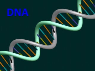
Each strand was in the shape of a helix The structure of DNA is now known to be a double helix. Watson & Crick also determined how the strands were connected
The Double-Helix Model • 1.accounted for Franklin’s X-ray pattern • 2. explains Chargaff’s rule of base pairing and how the two strands of DNA are held together.
DNA Deoxyribonucleic Acid de- away from
The Components of DNA • Nucleic acids are long, slightly acidic molecules originally identified in cell nuclei. • DNA is a nucleic acid made up of monomers called nucleotides joined into long strands or chains by covalent bonds.
Nucleotides are made of A phosphate group A nitrogenous base A 5-carbon sugar deoxyribose
Nitrogenous Bases and Covalent Bonds • The nucleotides in a strand of DNA are joined by covalentbonds formed between their sugar and phosphate groups.
Nitrogenous Bases and Covalent Bonds The nucleotides can be joined together in any order, meaning that any sequenceof bases is possible.
4 Different Nitrogenous bases Make Up DNA Adenine A Cytosine C Guanine G Thymine T
Nitrogenous Bases and Covalent Bonds • The nitrogenous bases stick out sideways from the nucleotide chain.
Forming the Double Helix Composed of sugar-phosphate strands ( two of them) The strands are held together by hydrogen bonds