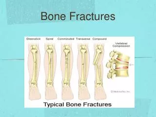
Trauma– Pelvic Fractures
920 likes | 5.04k Vues
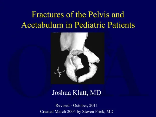
Trauma– Pelvic Fractures. Jay B. Baker, MD. Learning Objectives. Understand pathology of pelvic fractures Assess and work up trauma patients for pelvic fracture Treat the unstable pelvis Disposition pelvic fractures appropriately. Introduction.

Trauma– Pelvic Fractures
E N D
Presentation Transcript
Trauma– Pelvic Fractures Jay B. Baker, MD
Learning Objectives Understand pathology of pelvic fractures Assess and work up trauma patients for pelvic fracture Treat the unstable pelvis Disposition pelvic fractures appropriately
Introduction • Pelvic fractures are one of the few “talk and die” cases that can totally ruin your day • Associated with other severe injuries • Intraperitoneal– liver, spleen • Retroperitoneal– bladder, urethral • Vascular injuries including aortic rupture1 • Thus hypotension may be from a source other than the pelvis in blunt trauma
Sources of Blood Loss • Unstable pelvic fractures with hemorrhagic shock have four potential sources of blood loss– • Fractured bone surfaces • Pelvic venous plexus • Pelvic arterial injury • Up to 30% extrapelvicsources—chest, abdomen, multiple extremity, external1
Classification • Originally designed to predict bleeding risk, but not sensitive or specific enough3 • AP compression– “Open book” • Usually from “head on” trauma • Frequency is 15-20% • Lateral compression • Usually from “side on” trauma • Pelvic volume is compressed so life-threatening hemorrhage may be less likely • 60-70% • Vertical shear è major pelvis instability • Jumpers or “head on” MVC • 5-15% 1 • Combinations may occur, associated with major bleeding.
Assessment • Leg-length discrepancy or rotational deformity without fracture suggests unstable pelvis fracture • Motion may be detected by grasping iliac crests and pushing inward, then outward (aka compression-distraction maneuver) • Weingart recommends compression only4 • Once pelvic instability is demonstrated, no further maneuvers are necessary • Inspect flank, scrotum, and perianal area for bleeding including urethral meatus • Inspect for lacerations in perineum, vagina, rectum, or buttocks for open pelvic fractures • May defer vaginal speculum exam to OR • Palpate for high-riding prostate gland in males
Initial Imaging • AP xray of pelvis confirms clinical exam • FAST is specific for intra-abdominal injury but not for retroperitoneal bleeding • If positive, patient goes to OR for laparotomy • If negative, patient needs CT abd/pelvis • Some surgeons might perform supra-umbilical DPA to rule out intra-abdominal bleeding • Not infra- to avoid possible pelvic hematoma
CT Abdomen and Pelvis • Obtain CTAP in hemodynamically stable patient with clinical suspicion of intra-abdominal injury • Defer retrograde urethrogram (RUG) in severe pelvic trauma • Assessment for life-threatening injuries takes precedence • IV contrast for RUG may interfere with interpretation of pelvic angiogram or CTAP5
Management • Splint unstable pelvis fractures with simple techniques • Goal– Decrease pelvic volume prior to transfer and during resuscitation • Wrap a 12” sheet around the greater trochanters and tighten down6 • Commercially available pelvic splints • Other pelvis stabilizing devices e.g. ex-fix are not preferred due to time-intensive
Sample algorithm • Surgical consult • Pelvic splint • Evaluate for intraperitoneal gross blood • If (+), then dispo patient to the OR for laparotomy • If (-), then dispo to angiography1 • NB: Angioembolization may be performed concurrently with laparotomy
Controversy • Some centers perform pre-peritoneal packing first7 • May be performed simultaneously with laparotomy if necessary • A lot quicker when your surgeons are in house and your IR is still driving into the hospital • Some centers reserve angioembolizationfor non-coagulopathic patients who continue to bleed from a pelvic source despite pre-peritoneal packing
Summary Unstable pelvic fractures can bleed to death as a solo injury or in association with other severe injuries Unstable pelvic fractures must be splinted immediately Unstable pelvic fractures require prompt surgical management and may require angioembolization by interventional radiology
References • Fildes J et al. Advanced Trauma Life Support Student Course Manual (8th edition), American College of Surgeons 2008. • Nickson, Chris, “Trauma! Pelvic Fractures I.” Weblog entry. Life in the Fast Lane.com. April 23, 2012. http://lifeinthefastlane.com/2012/04/trauma-tribulation-027/ • Cullinane DC et al. Eastern Association for the Surgery of Trauma practice management guidelines for hemorrhage in pelvic fracture- update and systematic review. J Trauma 2011 Dec;71(6):1850-68. • Weingart, Scott, “Severe Pelvic Trauma.” Weblog entry. EMCrit blog: A discussion of the practice of ED critical care. April 30, 2012. http://emcrit.org/podcasts/severe-pelvic-trauma/ • Runyon MS. Blunt genitourinary trauma. In: UpToDate, Walls RM, Bachur RG (Ed), UpToDate, Waltham, MA, 2012. • McGonigal, Michael, “Compression Of The Fractured Pelvis With A Sheet.” Weblog entry. The Trauma Professional’s Blog. http://regionstraumapro.com/post/9038627564 • Burlew CC, Moore EE. Definitive management of severe pelvic fractures in adults. In: UpToDate, Frankel, HL (Ed), UpToDate, Waltham, MA, 2011.
Simulation Training Assessment Tool– Unstable Pelvic Fracture Jay B. Baker, MD
Simulation Training Assessment Tool (STAT)– Unstable Pelvic Fracture Date: Instructor(s): Learner(s): Learning Objectives: Recognize and treat pelvic fracture with hemodynamic instability Recognize intraabdominal bleeding with EFAST and treat with PRBCs Consult Trauma Surgery & Interventional Radiology for disposition ME = Meets Expectations; NI = Needs Improvement
Synopsis • This case assesses the learner’s management of an unstable pelvic fracture with hemoperitoneum from likely intraabdominal injury. • The setting is the ED resuscitation bay. The patient has a CC of back and pelvis pain after she was thrown from a horse and couldn’t walk due to pain. Her vitals are stable en route despite a brief SBP 85 that improved after NS 500. When the learners enter the room, they see Sim Man on a backboard and C collar w/o a monitor. The team leader (TL) must begin the 1° survey immediately and find patient is conscious, in pain, tachycardic, and mildly hypotensive. The TL must order fluid resuscitation and proceed to 2° survey while a team member performs the FAST- which is positive in Morrison’s Pouch. Secondary survey reveals TTP at LUQ, unstable pelvis, perineal ecchymosis, and positive gross blood at rectum. • The pelvic binder must be applied quickly after detection. The instructor will decrease the SBP to 80– this must be detected with repeat VS. If learner fails to reassess VS, instructor may cause the patient to lose consciousness. Improvisation may be required depending on learner’s decision making if this occurs. Transfusion with emergency release PRBCs should be initiated quickly with T&C for 4 units minimum regardless if H/H is obtained. Foley is contraindicated. If PRBCs are not ordered, patient may lose consciousness as above. • Patient’s BP will stabilize with transfusion. Due to hemoperitoneum, patient will dispo to the OR with general surgery consult. Interventional radiology must be considered for possible embolization. • If pain control is attempted, it should begin with low doses of short acting agents e.g. fentanyl 25 mcg. • The entire case should not take longer than 15 minutes. Personnel and roles • Instructor assesses learners with STAT, jockeys VS through two changes • Confederate nurse (possibly a Sr. resident) is helpful, not obstructive, doesn’t give away case Essential props and supply checklist • 3G Sim Man w/ C collar, back board, moulage: perinealbrusing and gross blood at rectum • Monitor, monitor leads, O2 mask, IV x 2, IVF • A pelvic binder that is stocked in your ED or a bed sheet • Simulated PRBCs Debriefing • Take notes with STAT to record impressions of learners’ performance with special attention to observed performance gaps. Use your notes to aid your debriefing. • Begin with Reaction phase– Allow learners to blow off steam, clarify facts of case, collect insight for Teaching Phase. 1-2 minutes. • Proceed into Teaching phase– Discuss each of the Critical Actions, mentioning each one in sequence. Proceed quickly where the learner performed well. Focus debriefing on areas where performance gaps were noted. 10-15 minutes. • Take time to teach how to apply a pelvic splint correctly if this is needed. • Summary phase– Ask learners to summarize briefly the following: • What did they do well that they will do with a real patient? Briefly augment with any additional observations. • What did they learn that they will do with a real patient? Briefly augment with any additional observations.
Images • Pelvic fx film • CXR • Lateral C spine • FAST x 4 windows w +MP
Labs • iSTAT • Na 140 • K 4.2 • Cl 100 • CO2 23 • BUN 18 • Cr 0.8 • Hct 24 • Hb 8.1 • AnGap 19