Download
1 / 16
230 likes | 583 Vues
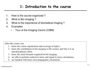
1: Introduction to the course. How is the course organized ? What is Bio-imaging ? What is the importance of biomedical imaging ? Examples Tour of the Imaging Centre (CIBM). After this course you know the course organization and coverage of topics;

E N D
1: Introduction to the course • How is the course organized ? • What is Bio-imaging ? • What is the importance of biomedical imaging ? • Examples • Tour of the Imaging Centre (CIBM) • After this course you • know the course organization and coverage of topics; • know the contribution of bio-imaging to life science and why it is an interdisciplinary effort. • know the main elements required for bio imaging; • are able to perform contrast to noise and signal to noise calculations; • are familiar with noise error propagation calculations
1-1. How is the course organized ? • Copies of the presentation • Will be provided on moodle (pdf) • ppsx : provided as link at lesson No. () • Please take notes during lecture without using “polycopies” • Exercises (Fri 15:15 CE 1): Handed out by assistant on day of lecture Available on moodle Solution of selected problems of prior week • Course web site(moodle, physics, master): • moodle.epfl.ch/course/view.php?id=250 • If we haven’t enrolled you yet: Enrollment key = bioimaging13 • If the lecture is unclear, holler ! • 90% chance you are not the only one • There are no stupid questions (I will take any question seriously) You can also approach me in the break I will be present in the exercises Your participation wanted! • If you miss a course … • The course given in 2011 was filmed • the link is provided on moodle • ppt animation do not show • youtube videos do not play fully
What is the content of this course ? • Links • Life science @ EPFL • Systems and signals • Image processing • Mathematical and computational models in biology • Physics • Neural networks and biological modeling • Classical electrodynamics
What supplemental reading/material is recommended ? • Other Text books • Zhang-Hee Cho, Joie J. Jones, Manbir Singh “Foundations of Medical Imaging” • William R. Hendee, E. RusselRitenour“Medical Imaging Physics” • Jerrold T. Bushberg, J. Anthony Seibert, Edwin M. Leidholt, John M. Boone “The Essential Physics of Medical Imaging” • I will provide pdf versions of the lecture on moodle • Handouts without your personal notes will not be complete. • To complete the Handouts • personal notes during course • incorporate insights gained during exos For a shorter text: Penelope Allisy-Roberts, Jerry Williams “Farr’s Physics for Medical Imaging” (200p., small, ~EUR 50) USD 30+ on amazon.com A lot of focus on simple x-ray (not covered in the course) • Course text: • Andrew Webb“Introduction to biomedical imaging”(250p. ~EUR 110, available as ebook at the library EPFL) • USD 60+ on amazon.com • Is more complete on MRI • Excellent reference text for later use
What do these images represent ? X-ray image of the hand Röntgen (1901) / Cormack & Hounsfield (1979) Electron microscopy of red blood cells Ruska (1986) Data: Rosalind Franklyn X-ray diffraction image of DNA crystals Watson, Crick, Wilkins (Nobel 1962) MRI of a head Lauterbur / Mansfield (2003)
1-2. What is Biomedical Imaging ? Definition of bio-imaging • What is measured (some useful definitions) • Image=nxm matrix of pixels Pixel = picture element • 3D image=kxnxm matrix of voxels Voxel = volume element What is Contrast ? Ability to distinguish tissue features against noise Contrast = difference in signal between tissues one wishes to distinguish In reality one needs to deal with Contrast-to-noise Localized measurement of a contrast generating biophysical effect in body/organ of living system Is there a free lunch for imaging ? Important: Contrast between voxels/pixels STOP! Resolution Sensitivity/Contrast In principle n,m,k can be unlimited…
What is the difference between signal-to-noise and contrast-to-noise ratio ? To discriminate two signals S1 and S2 we need more than just good signal to noise ratio. The ability to discriminate the two is assessed using the contrast to noise ratio Definition Contrast-to-noise ratio (CNR) S1 and S2 : two signals (or measurement variable) of two different tissues, s : standard deviation of their measurement (see left, assumed here to be identical and statistically independent) CNR provides a means to estimate the precision with which the signal S1 can be discriminated from S2 • To obtain good measurements (not only in imaging) we need good signal to noise ratio • Definition • Signal-to-noise ratio (SNR) • S: signal (or measurement variable) • s: standard deviation of its measurement (either determined experimentally (how?) or estimated quantitatively) • SNR provides a means to estimate the precision with which the signal S is measured It is possible to have excellent SNR but no CNR (when?)
How can we optimize SNR ? <eiej>=0, i≠j • It is possible to optimize SNR by performing N repeated measurements Si. • The precision of the average <S>=∑Si/N • depends on the square root law(4 measurements improve the precision by twofold): • Si=S+ei • where <ei2>=s2, <ei>=0. • S is the true signal (unknown) • <S>=∑Si/N=S+ ∑ ei/N This is well-known from statistics (SEM) results in increased measurement time
How can we optimize CNR ? • Optimizing contrast = choice of experimental parameters (e.g. protocol) to maximize the difference in two tissue signals S1 and S2. complex and empirical procedure some effects can be predicted/calculated, if the signal behavior can be modeled. =0 • Error propagation calculation • Let the signal S be a function S(k,t) • k is a tissue property (signal decay rate) • t an experimental parameter (such as time). • Approach: • Determine dS/dk • Find t0 where dS/dk is maximal by taking derivative rel. to t t0=1/k For an exponentially decaying signal, the optimal time of measurement is equal to 1/decay rate How critical is the choice of t0 ? Example: Maximum is where derivative with respect to t is zero t0
1-3. What is the importance of Bio-Imaging ? • Life Sciences are unthinkable without Bio-Imaging Assessment of biological processes with minimal perturbation of the system • Examples: • Humans, animals, cell/organ preparations • Modalities: • x-ray • computed tomography • positron emission tomography • magnetic resonance • ultrasound • electrical imaging (EEG, MEG) • optical imaging Development of Bio-Imaging capabilities, modalities and effects … unthinkable without physics
What are essential ingredients of bio-imaging ? • Life Sciences • Physics • Engineering/Good instrumentation • Mathematics • Chemistry Bio- & organic chemistry Neuroscience Electrodynamics Quantum mechanics Thermodynamics Classical Mechanics Electrical Mechanical Cancer Multi-disciplinarity is important ! Biomedical engineering Mathematics
What is the perfect imaging modality ? • Easy to use • Portable • Highly sensitive/good contrast • Does this exist ? In reality, every imaging method/modality has its strengths and limitations In this course you will learn to appreciate these and the reasons behind
1-4. ExamplesAutoradiography Autoradiography of a brain slice Autoradiography of a gel Autoradiography of a monkey brain (visual cortex)
What are the distinct advantages of Bio-imaging compared to tissue analysis ? • Imaging advantages • relative to histology or invasive tissue analysis • Rapid acquisition of the information • Non-destructive, i.e. minimal perturbation • In situ or in vivo • Repetitive (longitudinal) studies possible Mice subjected to 30 min of stroke assessed using MRI before and 3-24h after Ultrasound of mouse heart Histology:Tissue is fixed, cut into slices, then subjected to a dye. The resulting sections are then analyzed.
Examples: Biomedical Imaginghttp://nobelprize.org/educational_games/physics/imaginglife/narratives.html Metastasis localization (PET) 3D rendering of tumor for surgical planning (MRI) fMRI of whole brain activation
What’s next today ? 1. Tour of CIBM If you would like to volunteer for a brief (15min) scan of your brain … let us know 2. Exercise (~45min)