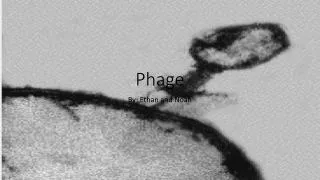
What is a Phage?
What is a Phage?. Phage Landing and Infusion of Viral DNA. Phage Lifecycle: Lytic vs Lysogenic. Lytic 1) Virus infuses DNA into host cell 2) Virus quickly hijacks cellular replication machinery 3) Replicates it’s own viral DNA First reverse translation if retrovirus


What is a Phage?
E N D
Presentation Transcript
Phage Lifecycle: Lytic vs Lysogenic • Lytic • 1) Virus infuses DNA into host cell • 2) Virus quickly hijacks cellular replication machinery • 3) Replicates it’s own viral DNA • First reverse translation if retrovirus • 4) Cell wall bursts releasing newly synthesized viral phages into surrounding medium • Lytic • 1) Virus infuses DNA into host cell • 2) Virus quickly hijacks cellular replication machinery • 3) Replicates it’s own viral DNA • First reverse translation if retrovirus • 4) Cell wall bursts releasing newly synthesized viral phages into surrounding medium • Lysogenic • 1) Virus infuses DNA into host cell • 2) Viral DNA incorporates itself into host DNA, and exists dormantly • 3) Stress or stimulation induces virus DNA to be come active • 4) Lytic life cycle begins
Bacterial lysis via Lysin • Lysin • Viral enzyme that facilitate bacterial host lysis • Hydrolyze bonds essential for peptidoglycan integrity • Amidases are a class Lysins : • Well conserved N-terminal catalytic domain • Highly divergent C-terminal binding domain • high binding constant (KA = 3.6 *108, similar to Ab binding) • recognizes species specific carbohydrates that are essential for viability • Therefore evolution of resistance to lysins unlikely
PlyG Identification • Screened expression libraries for possible lysing agents of RSVF-1 • All lytic clones identified contained a 702 bp ORF encoding a homologue of N-acetylmuramoyl-L-alanine amidase (~27kDa) • class of phage lysins • Homology restricted to catalytic N-terminal portion of protein • Table 1: • high specificity and lytic activity for all B. anthracis strains • similar specificity and activity for B. cereus RSVF-1 • RSVF-1 chosen for remainder of experiments probably for safety • note: titer measurement analog to agglutination titer; Immunology text (p.245) • Here titer refers to lysis rather than agglutination
Assay for Lytic Phage • Plaque assay - Lytic phage are enumerated by a plaque assay. A plaque is a clear area which results from the lysis of bacteria (Figure 4). Each plaque arises from a single infectious phage. The infectious particle that gives rise to a plaque is called a pfu (plaque forming unit).
Fig. 1 • Conserved N-terminal catalytic domain • Divergent C-terminal binding domain
Fig. 1 Circle represents void of bacterial cells as a result of PlyG lysis (0.5 U of PlyG added to plate)
(# Viable Bacteria after buffer treatment) (# Viable Bacteria after 20U PlyG treatment) Fold Killing = Fig. 2 20U PlyG
Fig. 2 2U PlyG Buffer CFU correlation to A600 2U PlyG • 17000 fold decrease w/in 20 sec • near sterilization at 2 minutes • RLU • relative light units • signal emitted by Luciferin in presence of ATP released by lysed bacterial cells • CFU • Colony forming units (part (b) inset due to “strong detergent mix”?)
Fig. 3 Phase contrast microscopy No PlyG added 1 min after 5U added 15 min after 5U added 1 min after 5U added 10 min after 5U added Transmission Electron Micrographs (TEM)
Fig. 4 In Vivo mouse (BALB/c) infection/survival model No B. anthracis added 150U PlyG added 50U PlyG added No PlyG added
Fig. 5 PlyG mediated spore killing and detection 10U PlyG 5 min after 2U PlyG added (b) & (c) 60 min after 2U PlyG added D-alanine L-alanine D-alanine inhibits spore germination L-alanine induces spore germination • RLU • relative light units • signal emitted by Luciferin in presence of ATP released by lysed bacterial cells • CFU • Colony forming units (part (b) inset due to “strong detergent mix”?)
Conclusion • PlyG: specific B. anthracis lysin identified and purified • Activity and specificity verified in vitro • Luciferin activity due to ATP release from lysed cells is a specific assay of PlyG activity • PlyG rapidly kills g-sensitive bacteria in vivo • Resistance to PlyG not observed • even under induced mutagenesis conditions which caused >1000 fold increases in acquired antibiotic resistance as compared to uninduced mutagenesis. • g phage resistance observed to evolve • Therefore PlyG likely to target essential cell-wall molecules • And therefore intrinsic PlyG resistance is unlikely • Only germinating spores are sensitive to PlyG • RLU of only 100 RSVF-1 spores detected with handheld luminometer represents potential detection techniques of “battlefield and home” use of B.anthracis
PlyG Lysin Specificity • Table 1: • high specificity and lytic activity for all B. anthracis strains • similar specificity and activity for B. cereus RSVF-1 • RSVF-1 chosen for remainder of experiments probably for safety • note: titer measurement analog to agglutination titer; Immunology text (p.245) • Here titer refers to lysis rather than agglutination • Figure 1: • next slide
PHAGE MULTIPLICATION CYCLE • A. Lytic or Virulent Phages • 1. Definition - Lytic or virulent phages are phages which can only multiply on bacteria and kill the cell by lysis at the end of the life cycle. • 2. Life cycle - The life cycle of a lytic phage is illustrated in Figure 3 . • a. Eclipse period - During the eclipse phase, no infectious phage particles can be found either inside or outside the bacterial cell. The phage nucleic acid takes over the host biosynthetic machinery and phage specified m-RNA's and proteins are made. There is an orderly expression of phage directed macromolecular synthesis, just as one sees in animal virus infections. Early m-RNA's code for early proteins which are needed for phage DNA synthesis and for shutting off host DNA, RNA and protein biosynthesis. In some cases the early proteins actually degrade the host chromosome. After phage DNA is made late m-RNA's and late proteins are made. The late proteins are the structural proteins that comprise the phage as well as the proteins needed for lysis of the bacterial cell. • b. Intracellular Accumulation Phase - In this phase the nucleic acid and structural proteins that have been made are assembled and infectious phage particles accumulate within the cell. • c. Lysis and Release Phase - After a while the bacteria begin to lyse due to the accumulation of the phage lysis protein and intracellular phage are released into the medium. The number of particles released per infected bacteria may be as high as 1000.