Download
1 / 22
220 likes | 443 Vues
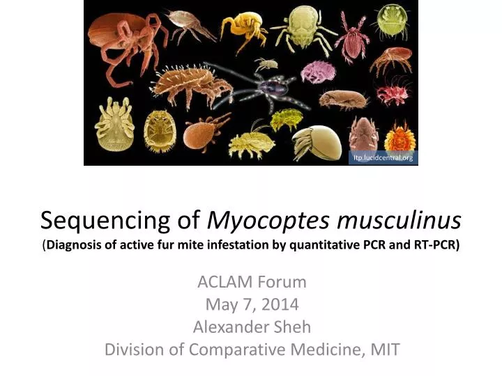
Sequencing of Myocoptes musculinus ( Diagnosis of active fur mite infestation by quantitative PCR and RT-PCR). Itp.lucidcentral.org. ACLAM Forum May 7, 2014 Alexander Sheh Division of Comparative Medicine, MIT. Common rodent fur mites Myocoptes musculinus Myobia musculi

E N D
Sequencing of Myocoptesmusculinus(Diagnosis of active fur mite infestation by quantitative PCR and RT-PCR) Itp.lucidcentral.org ACLAM Forum May 7, 2014 Alexander Sheh Division of Comparative Medicine, MIT
Common rodent fur mites Myocoptesmusculinus Myobiamusculi Radfordiaaffinisand ensifera Other mites Demodexmusculi, arvicolaeand flagellurus Psorergatessimplex Mesostigmatic mites Laelapsechidnina(spiny rat mite) Ornithonyssusbacoti(tropical rat mite) Liponyssoidessanguineus(house mouse mite) Dermatophagoidesfarinaeand pteronyssus(house dust mites) Mites
M. musculinusand M. musculi • Most common fur mites in lab mice. • Nonburrowing, feeding on skin secretion and interstitial fluid • Often present in mixed infections • Myobia~ head/shoulder pelage • Myocoptes~ inguinal, ventral abdomen and dorsum • Transmission is direct (not through bedding) and requires hair shafts http://www.idexxbioresearch.com
Effects of mite infestation • General health complications • None • Ruffled fur to alopecia to erythema to pruritus to ulcerative dermatitis • Strain dependent • Decreased life span, body weight and reproductive indices • Chronic infestations -> dermatitis may provoke secondary amyloidosis • Self-trauma (secondary bacterial infections) • Research concerns • Immunological research • Behavioral research • Endocrine research • Toxicology research
Treatment • Drugs (Ivermectin, Selemectin, Permethrins, Chlorpyrifos, Dichlorvos, Moxidectin, and others) • Injection, in water, in feed, spot on treatment, bedding, and nestlet soak. • Varied results with drug treatment based on species and treatment method • Rederivationmay be needed to clean up a colony • Prevention is preferred over treatment mosup.com
Diagnostic methods • Methods • Direct exam of pelage • Cellophane tape testing • Hair plucks • Skin scraping • PCR • Typically, detection of fur mites is a visual process and poses difficulties in terms of throughput, sampling and personnel requirements. http://www.idexxbioresearch.com
Diagnostics by PCR • Charles River and IDEXX RADIL offer fur mite PCR assays targeting rRNA genes. • PCR increases throughput/sampling and reduces hands on time with good specificity and sensitivity.
RT-PCR: How do you know you eradicated mites? • Due to DNA’s stability, residual mite tissue on treated mice may be a potential source of PCR false positives (RicartArbona et al. 2010). • While DNA dominates the forensic sciences, understanding RNA and its degradation may offer insights into cause of death, the age of wounds/injuries and the post-mortem interval(Bauer 2007). • 16S rRNA detection from Chlamydia pneumoniae was better associated to active infection than detection of specific antigens(Meijer et al., 2000). • Can we use RNA degradation to complement DNA based assays?
Developing a RT-PCR assay for mites • Sequence rRNA and mitochondria • Myocoptesmusculinus • Myobiamusculi • Radfordiaaffinis • Develop and test specific primers
Available fur mite sequences on NCBI • 935 bp sequenceMyocoptesmusculinus18s ribosomal RNA gene • Closely related Myocoptesjaponensis has available 18S and 28S sequences • Myobiamusculi 18S, ITS, 5.8S, ITS, partial 28s rRNA(S.Compton (2011) and S. Feldman (2011)). • No mitochondrial sequences available
Phylum Arthropoda, Class Arachnida The common murine fur mites diverge at superorder Acariformes. The genera Myobiaand Radfordiaare both in the Myobiidae family. The genus Myocoptesis within the superfamily Sarcoptoidea and the family Myocoptidae. Myocoptesmusculinusand Dermatophagoidesspp. diverge at suborder Psoroptidia. • Superorder Parasitormes • Laelapsechidnina, Ornithonyssusbacoti, Liponyssoidessanguineus • Order Trombidiformes • Myobiamusculi, Radfordiaspp., Psorergates simplex, Demodexmusculiand other (Tetranychusurticae– spider mites) • Order Sarcoptiformes • Myocoptesmusculinus, Dermatophagoidesspp. and other (Sarcoptesscabiei- scabies) Adapted from www.tolweb.org
Processing Myocoptessamples • Mites obtained from an experimentally infested colony at MSKCC (Dr. Neil Lipman). • Genomic DNA was extracted from fur plucks from individual mice using the QIAamp® DNA Micro kit (Qiagen) using carrier RNA. DNA was pooled.
Primer selection • Three pairs of rRNA primers based on Myocoptesjaponensissequences. • Mitochondrial primers based on complete mitochondrion sequences for Dermatophagoidespteronyssus(EU884425) and Dermatophagoidesfarinae(NC_013184). • More nonspecific bands! rRNA PCR
MiSeq Sequencing • Gel extracted PCR amplicons were pooled into rRNA or mitochondria samples • Amplicons were sonicated, size-selected and ligated to sequencing adapters. • Samples were ligated and amplified on flow cell for sequencing. Illumina
MiSeq Sequencing • 150 bp paired end reads were sequenced and assembled by Velvet. • Contigs analyzed by Geneious Illumina
Ribosomal RNA alignment • Generated a 6.35Kb contig from assembly of rRNA sample reads • Compared to Myobiamusculi, Dermatophagoidesspp., MyocoptesjaponensiandMyocoptesmusculinus, with Musmusculusas negative control (77%) • Data from Charles River showed a 92% homology between Myobiamusculiand Myocoptesmusculinus(Henderson and Perkins seminar) Myobia 84.6% D. farinae 92.9% Increased taxonomic similarity M. japonensis 98.9% M. musculinus 99.9%
Phylogenetic analysis of subclass Acari 18S rRNA sequences • Sanity check – created a phylogenetic tree using 18S sequences from 223 species from the subclass Acari Trombidiformes, incl. Myobia Parasitiformes, Trombidiformes, Sarcoptiformes Myocoptesmusculinus Sarcoptiformes
Mitochondrial genome alignment • More difficulties with mitochondrial genome • Have generated 5.2Kb out of ~14Kb • Blue denotes new sequences matching reference • Green and red denote reference genome • Purple denotes PCR products • 71.1-72.9% homology to D. farinaeand pteronyssus, respectively. • Sequenced cox1-3, atp6, atp8, and cytB genes
Future directions • Sequence rest of Myocoptesmusculinusmitochondria, and mitochondria and rRNA of Myobiamusculiand Radfordiaaffinis • Design primers and test on PCR products and T7-generated ssRNA under diverse degradation conditions. • Obtain frozen samples from Ivermectin-treated and untreated mice
Acknowledgements • ACLAM • James G. Fox • Mark Whary • DCM postdocs • Laura Cacciopo, Courtnye Jackson, Courtney Ek • Neil Lipman - MSKCC • BioMicro Center
References • Pathology of Laboratory Rodents and Rabbits. Percy and Barthold, eds. 2007, 3rd ed. • The Mouse in Biomedical Research, 2nd Edition. Volume II, Diseases. 2007. Fox et al., eds.. • Flynn’s Parasites of Laboratory Animals. Second Edition. 2007. Baker, ed. • RicartArbonaet al. Treatment and eradication of murine fur mites: I. Toxicologic evaluation of ivermectin-compounded feed. JAALAS 2010;49(5):564-70 • RicartArbonaet al. Treatment and Eradication of Murine Fur Mites: III. Treatment of a Large Mouse Colony with Ivermectin-Compounded Feed. JAALAS 2010;49(5):633-7. • RicartArbonaet al. Treatment and Eradication of Murine Fur Mites: II. Diagnostic Considerations. JAALAS 2010;49(5):583-7. • Lindstrom et al. Soiled Bedding Sentinels for the Detection of Fur Mites in Mice. JAALAS 50(1)p54-60. • Mooket al. Use of selamectin and moxidectin in the treatment of mouse fur mites. JAALAS 2008 May;47(3):20-4. • Burdett et al. Evaluation of five treatment regimens and five diagnostic methods for murine mites. JAALAS 1997 Mar;36(2):73-6. • Carty, A. Opportunistic infections of mice and rats: Jacoby and Lindsey revisited. ILAR Journal, 49(3), 2008. 272-276. • Cliffordand Watson. Old enemies, still with us after all these years. ILAR Journal, 49(3), 2008. 291-302. • Watson. New building, old parasite: Mesostigmatid mites-an ever-present threat to barrier rodent facilities. ILAR Journal, 49(3), 2008. 303-309. • http://tolweb.org/tree/ • Hill et al. ContempTop Lab Anim Sci. 1999 Nov;38(6):13-18. Demodexmusculi in the Skin of Transgenic Mice. • Dermauw et al. 2009 BMC GENOMICS. 10. The complete mitochondrial genome of the house dust mite Dermatophagoidespteronyssinus (Trouessart): a novel gene arrangement among arthropods • http://www.criver.com/about-us/eureka/wp-content/uploads/Fur-mite-PCR-AALAS-KSH-talk.pdf • http://www.idexxbioresearch.com/radil/userfiles/download_files/MitesFlyer_US_Rev062012.pdf • Weiss et al. Comparison of a Fur Mite PCR Assay and the Tape Test for Initial and Posttreatment Diagnosis during a Natural Infection JAALAS Volume 51, Number 5, September 2012 , pp. 574-578(5) • Rice et al. Evaluation of Diagnostic Methods for MyocoptesmusculinusAccording to Age and Treatment Status of Mice (Musmusculus). JAALAS Volume 52, Number 6, November 2013 , pp. 773-781(9) • Karlsson, EM et al. Combined Evaluation of Commonly Used Techniques, Including PCR, for Diagnosis of Mouse Fur Mites. JAALAS, Volume 53, Number 1, January 2014 , pp. 69-73(5) • Bauer. RNA in forensic science. Forensic Science International: Genetics Volume 1, Issue 1, March 2007, Pages 69–74 • Meijer et al. Chlamydia pneumoniae in vitro and in vivo; a critical evaluation of in situ detection methods J. Clin. Pathol., 53 (2000), pp. 904–910. • Tolker-Nielsen et al. Effects of stress treatments on the detection of Salmonella typhimuriumby in situ hybridization Int. J. Food Microbiol., 33 (1997), pp. 251–258.