Download
1 / 30
340 likes | 944 Vues
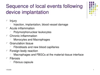
Sequence of local events following device implantation. Injury Injection, implantation, blood vessel damage Acute inflammation Polymorphonuclear leukocytes Chronic inflammation Monocytes and Macrophages Granulation tissue Fibroblasts and new blood capillaries Foreign body reaction

E N D
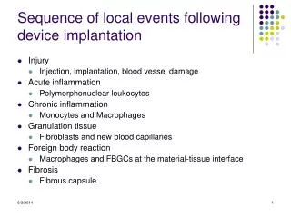
Sequence of local events following device implantation • Injury • Injection, implantation, blood vessel damage • Acute inflammation • Polymorphonuclear leukocytes • Chronic inflammation • Monocytes and Macrophages • Granulation tissue • Fibroblasts and new blood capillaries • Foreign body reaction • Macrophages and FBGCs at the material-tissue interface • Fibrosis • Fibrous capsule
Hemostasis: Vasoconstriction & Plug Formation Figure 16-12: Platelet plug formation
Hemostasis The process of blood clotting and then the subsequent dissolution of the clot, following repair of the injured tissue. Composed of 4 major events that occur in a set order following the loss of vascular integrity: 1. vascular constriction. This limits the flow of blood to the area of injury. 2. platelets become activated by thrombin and aggregate at the site of injury, forming a temporary, loose platelet plug. The protein fibrinogen is primarily responsible for stimulating platelet clumping. Platelets clump by binding to collagen that becomes exposed following rupture of the endothelial lining of vessels.
Hemostasis (continued) Upon activation, platelets release ADP and TXA2 (which activate additional platelets), serotonin, phospholipids, lipoproteins, and other proteins important for the coagulation cascade. In addition to induced secretion, activated platelets change their shape to accommodate the formation of the plug. 3. To insure stability of the initially loose platelet plug, a fibrin mesh (also called the clot) forms and entraps the plug. 4. Finally, the clot must be dissolved in order for normal blood flow to resume following tissue repair. The dissolution of the clot occurs through the action of plasmin.
Platelet Activation Platelets bind to matrix and spread to cover the damaged surface; aggregation to form temporary plug; • Initiates the wound healing process through the secretion of soluble small molecules from cytoplasmic granules called growth factors and cytokines (Platelet derived growth factor (PDGF), Fibronectin, von Willebron Factor and Transforming Growth Factor-beta (TGF-b); • These substances are sticky and bind to matrix, chemotactic (draw cells up the concentration gradient through migration) and /or mitogenic agents for leukocytes, endothelial cells and fibroblasts;
Hemostasis: Vasoconstriction & Plug Formation Figure 16-12: Platelet plug formation
Fibrin Clot Formation-Thrombogenesis Two principle pathways: converge on the same end product-fibrinogen fibrin Intrinsic pathway: clot in response to an abnormal vessel wall superficial injury in the absence of tissue injury Extrinsic pathway: clot formation in response to tissue injury , actual breakage of blood vessels. Both pathways are complex and involve numerous proteolytic enzymes called clotting factors.
Hemostasis The intrinsic pathway is the longer, slower pathway when compared to the extrinsic pathway. The intrinsic pathway can take between a few seconds or even minutes to produce Factor X. The extrinsic pathway reacts almost instantaneously by producing Factor X. The benefit of the intrinsic pathway is that more Factor X is produced. The extrinsic pathway's main function is to augment the intrinsic pathway by slowing the flow of blood outside the vessel by producing little Factor X, but quickly. The extrinsic pathway completes the clot and allows for the blood vessel to be repaired
Hemostasis: Coagulation & Clot Stabilization • Prothrombin • Ca++ • Fibrinogen • Fibrin • Polymerization Figure 16-13: The coagulation cascade
Factor Trivial Name(s) Pathway Characteristic I Fibrinogen Both - II Prothrombin Both Contains N-term. gla segment III Tissue Factor Extrinsic - IV Calcium Both - V Proaccelerin, labile factor, accelerator (Ac-) globulin Both Protein cofactor VI (Va) Accelerin - This is Va, redundant to Factor V VII Proconvertin, serum prothrombin conversion accelerator (SPCA), cothromboplastin Extrinsic Endopeptidase with gla residues VIII Antihemophiliac factor A, antihemophilic globulin (AHG) Intrinsic Protein cofactor IX Christmas Factor, antihemophilic factor B,plasma thromboplastin component (PTC) Intrinsic Endopeptidase with gla residues X Stuart-Prower Factor Both Endopeptidase with gla residues XI Plasma thromboplastin antecedent (PTA) Intrinsic Endopeptidase XII Hageman Factor Intrinsic Endopeptidase XIII Protransglutamidase, fibrin stabilizing factor (FSF), fibrinoligase Both Transpeptidase Primary Factors
Dissolving the Clot and Anticoagulants Figure 16-14: Coagulation and fibrinolysis
Complement Activation • Blood-materials interactions-protein adsorption; • The Complement system is a complex cascade involving approximately 30 glycoproteins present in serum as well as cell surface receptors; • Activation of the inflammation and immune related function.
Cytokines and Growth Factors • Autocrine (affect function of the cell that releases it) • Paracrine (affect the function of adjacent or nearby cells of the same or different phenotype)
TGF-b Chemoattractant for monocytes and fibroblasts • Pro-fibrogenic • stimulates fibroblast proliferation • Stimulates fibroblasts to secrete matrix (collagen, fibronectin, and glycosaminoglycans) and therefore aids in the development of wound strength • Stimulates angiogenesis (new blood vessel development)
Cellular Terminology: • granulocyte: any blood cell containing specific granules (e.g. neutrophils, eosinophils, basophils) • leukocyte: a colorless blood cell capable of ameboid movement (e.g. lymphocytes, monocytes, granulocytes) • macrophage: large phagocytic mononuclear cell
Clinical Signs of Inflammation: • redness (rubor), swelling (tumor), pain (dolor), heat (calor) Why rubor? erythrocytes Why swelling? Permeability: • pressure difference between capillary and external tissue bed • endothelium is tight permits very slow flow of water and small molecules into surrounding tissue NORMALLY: lymphatic vessels drain away this fluid maintaining constant tissue volume INFLAMMATION: permeability increases and larger molecules move into the tissue • increased fluid influx not promptly balanced by the lymphatic system • swelling (tumor)
Acute Inflammation Lasts from minutes to days depending on the injury Initial stages: • rapid dilation of local capillaries • increase in the permeability of their endothelial cell linings Dilation? • foreign protein or material coagulation factor (factor XII) kinins dilation and endothelial permeation Dilation leads to an increase in blood entry into the capillary beds • loss of plasma through the capillary walls • platelets and erythrocytes become sticky • blood flow slower and sludgy
Neutrophil (a granulocyte) First Cells to Appear at Injury Site • stick to capillary endothelium, penetrate between the endothelial cells and move into the surrounding damaged tissue; • neutrophil emigration (diapedisis) begins minutes to hours after insult and may continue for as long as 24h; • neutrophil activates when engages foreign particle such as a damaged cell, pathogen, damaged matrix, or a biomaterial; and, they • release interleukin-1 and tumor necrosis factor (TNF-alpha) called proinflammatory cytokines because they recruit monocytes to the injury site.
The Wound Healing Continuum • Initiation by mechanical injury/damage to vasculature • Blood coagulation-clot formation • Platelet activation and degranulation • Inflammation-edema • Removal of damaged matrix and necrotic cell components • Cell proliferation and recruitment including endothelial, epithelial, stromal and inflammatory cells • Continued removal of matrix • Angiogenesis • Matrix synthesis and deposition • Epithelialization and wound contraction • Decrease in cellularity-apoptotic pathway • Tissue remodeling-elastin synthesis