Advancements in CT Technology: A Comprehensive Overview from QIBA 2010
In a poll of 225 top general internists, CT and MRI were deemed the most significant medical advancements in the last 50 years, surpassing critical therapies like coronary angioplasty. This presentation outlines the evolution of CT technology from its prototype in 1972 to modern multi-detector systems, showcasing enhancements in spatial and temporal resolution, noise reduction, and radiation dose reduction. Additionally, it discusses the shift towards quantitative imaging, addressing structural issues and standardization challenges in the field, essential for clinical application and multi-vendor reproducibility.

Advancements in CT Technology: A Comprehensive Overview from QIBA 2010
E N D
Presentation Transcript
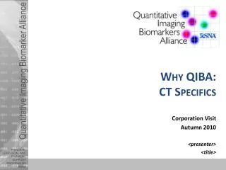
Why QIBA: CT Specifics Corporation Visit Autumn 2010 <presenter> <title>
In a poll of 225 top general internists, CT and MRI were judged to be the most important medical advances in the last 50 years, beating out life-saving therapies such as coronary angioplasty and ACE inhibitors. Fuchs VR, Sox HC Jr. Physicians' views of the relative importance of thirty medical innovations. Health Aff, 2001. 20(5): p. 30-42. Image courtesy of Toshiba Why QIBA: CT Specifics
Image courtesy of Siemens The Long and Proud History of Innovation of CT • 1972: Prototype CT • Several hrs per slice acquisition; days for reconstruction • 1974: 1st Generation CT • 2.5 min/slice • 1976: Whole-body CT • 5 sec/slice • 1989: Helical/Spiral CT • 0.3 sec/slice; 40 sec for entire chest (40cm Z-axis) • 1998: 4-row MDCT • 10 sec for entire chest • 2002: 16-row MDCT • 8 sec for entire chest • 2004: 64-row MDCT • 5 sec for entire chest • 20010 and beyond: ? Why QIBA: CT Specifics
Since 2004: Spatial Resolution up to 2x higher 3mm Stent Images courtesy of General Electric Non HDCT HDCT Why QIBA: CT Specifics
Temporal resolution over 2x faster Low Temporal Resolution High Temporal Resolution Why QIBA: CT Specifics
Stair-step Artifacts up to 80% Less Narrow Volume – 64 detector row CT Wide Volume – 320 detector row CT Images courtesy of Toshiba Why QIBA: CT Specifics
Image Noise up to 50% Less 60 mAs 60 mAs Images courtesy of General Electric Conventional Reconstruction HD Reconstruction Why QIBA: CT Specifics
Radiation Dose Reduction(one example of many promising approaches) Image courtesy of Siemens Why QIBA: CT Specifics
Multi-energy and Spectral CTTowards resolving multiple energies and materials 34 keV 51 keV Iodine (green) + Gd (red) • Automatic separation of iodine contrast and bone in a mouse Image courtesy of Philips Schlomka et al. Phys Med Biol (2008). Why QIBA: CT Specifics
Quantitative CT Imaging • The HU has always been “quantitative”, • Technical Advances will help us move from “qualitative image” to “quantitative image” or measurement • Longitudinal quantification used to assess patient response to therapy Why QIBA: CT Specifics
Picture Slide to Demonstrate Effects of Technology Advancement • (Jim Mulshine’s suggestion of Reeves slides here) • Lung lesion with • 10 mm thick images • 5 mm thick images • 2.5 mm thick images • 1 mm thick images • Volumetric representation of each • Showing how blurry and “artifacty”the 10 mm based image is and how clear the 1mm is. Why QIBA: CT Specifics
Movement to Quantitative Imaging • Series of 3 or 4 slides (primarily with pictures) showing: • Simple phantom lesion (from 1A?) measured twice (either same reader twice or two readers) • Complex phantom lesion measured twice • Show diameters • Show volumetric contours • Simple patient lesion (1B) measured twice • Complex patient lesion (1B) measured twice Why QIBA: CT Specifics
Use a better example, but ~ like this lesion Reader 1 contour (includes sliver) Reader 2 contour (excludes sliver) Even with Exquisite images, still uncertainty about what is and isn’t part of a lesion. This leads to uncertainty in measurements, even with experts. Why QIBA: CT Specifics
Structural Issues Currently Impede Realization of the Opportunity Efforts by individual manufacturers to qualify quantitative imaging applications: • Are more costly, and • Run over longer time periods… …than the business model of device and software manufacturers generally support. Endpoint qualification cost Assay validation Development These issues are exacerbated by lack of clarity in regulatory and reimbursement policy which increase the risk while decreasing the incentive time Even when individual companies do these steps, community need for standards required to address multi-vendor reproducibility are not accounted for. June 2010 Buckler Biomedical LLC 14 Why QIBA: CT Specifics
What we need to Meet the Opportunity • Need domain experts: • Problem assessment: which problem could be solved by image analysis algorithms • Usability: are the developed algorithms user friendly and really useful for the experts • Feedback from opinion leaders • Market opportunities • Have to evaluate their hardware/software: • Standardized image database with annotations • Definitions of standards • Evaluation: • Phantom data • Clinical trials • Clinical studies • First users (domain expert) • Cooperation with hardware vendors, because: • DICOM and other standards • Different vendors / Different image data quality • Interfaces with the hardware of different hardware vendors • Different vendors /Different image data acquisition filters • Different Vendors / Different data representation algorithms and hardware (screen) • Negotiations etc. with Regulatory (compliance FDA and other international regulatory)
We Have Three Choices Don’t Do It Do it Individually Do it Together Accept Higher Costs and Lower Reproducibility Seek the Win-Win Structure that Rewards Participants Accept Lower Utilization and Reimbursement Lowest utilization and reimbursement Localized utilization and reimbursement Highest utilization and reimbursement June 2010 Buckler Biomedical LLC 16 Why QIBA: CT Specifics
The Need • Need the appropriate Networks (Groups) in order to achieve the previous goals • Because of the • power of cooperation • Exchange experience • Exchange knowledge • Work with the regulatory (like FDA…) • State of the art knowledge • Etc.
How We Will Succeed Result: Widely Available, High Performance, Quantitative Imaging Make it actionable for engineering and R&D, addressing both design and use Make it familiar to marketing and give them a product, not just a cost Provide a regulatory pathway that works in the business model Imaging Science, Metrology, and Biostatistics June 2010 Buckler Biomedical LLC 18 Why QIBA: CT Specifics
QIBA Overcomes Obstacles to Enable the Opportunity Clinical Context Ground work Quant ImagingProfiles Profile Claims Profile Details With an IHE -like certification reputation June 2010 Buckler Biomedical LLC 19 Why QIBA: CT Specifics
Profiles are actionable for both Marketing and R&D PRODUCT CREATION PROCESS (PCP) QIBA PROFILE I. CLINICAL CONTEXT II. CLAIMS III. PROFILE DETAILS IV. COMPLIANCE SECTION V. ACKNOWLEDGEMENTS Customer Requirements Specification (CRS) System Requirements Specification (SRS) Verification Plan and Protocol Participation and visibility June 2010 Buckler Biomedical LLC 20 Why QIBA: CT Specifics
Example: Lung Cancer Make drug development more efective: • Faster (Window trials—quantitative endpoint);Cheaper (Adaptive Bayesian Design, two to three weeks of drug exposure);Better (Phantom calibration, standardize method, open source reference tools, defined molecular targets, tailored delivery systems) • Tighter (variance), lighter (dose), standardized (protocol/profile) Make care more personalized to patient: • Clinically proven detection and longitudinal quantification • Quantitiative CT measures incorporated into adaptive therapy / monitoring Why QIBA: CT Specifics
Example: Emphysema and COPD • Chronic obstructive pulmonary disease (COPD) is a progressive lung disease which include emphysema, chronic bronchitis, refractory (irreversible) asthma, and severe bronchiectasis. • 12 million adults have COPD, and another 12 million are undiagnosed or developing COPD. It is the fourth leading cause of death in the U.S., accounting for 126,129 deaths in 2003. • COPD does not have a cure, but treatments are currently available to help individuals manage their symptoms of COPD Why QIBA: CT Specifics
Utility of CT evaluation of COPD • A. Dirksen et al., "A randomized clinical trial of alpha(1)-antitrypsin augmentation therapy," Am J Respir Crit Care Med 160, 1468-1472 (1999). • “We conclude that lung density measurements by CT may facilitate future randomized clinical trials of investigational drugs for a disease in which little progress in therapy has been made in the past 30 yr.” • In 2009: ”…CT is a more sensitive outcome measure of emphysema-modifying therapy than physiology and health status, and demonstrates a trend of treatment benefit from alpha(1)-AT augmentation.” • Present limitations of CT evaluation of emphysema: • Density measurements vary because level of inspiration varies. • There are inconsistencies between lung density measurements made using different vendor scanners, particularly using contemporary scanners. • May be associated with differences in CT number scale. Protocol Profile “Target” Profile “Ideal” Why QIBA: CT Specifics
QIBA: Active in Several Aspects Analyzing/Creating Data to Inform Profiles QIBA PROFILE I. CLINICAL CONTEXT II. CLAIMS III. PROFILE DETAILS IV. COMPLIANCE SECTION V. ACKNOWLEDGEMENTS QIBA Experiments and Groundwork • Analyzing: • Effects of Measurement Methods • 1D, 2D, 3D • Effects of Slice thickness • Phantoms • Apply to Patient Images • e.g. Coffee Break Experiment • Standardization across scanners June 2010 Buckler Biomedical LLC 24 Why QIBA: CT Specifics
QIBA: Active in Several Aspects UPICT Profiles (Target Concept) Acceptable QIBA PROFILE I. CLINICAL CONTEXT II. CLAIMS III. PROFILE DETAILS IV. COMPLIANCE SECTION V. ACKNOWLEDGEMENTS Target Ideal Determining Which Parameters (Slice Thickness, Recon Algorithm, etc.) Affect Measurement Variability June 2010 Buckler Biomedical LLC 25 Why QIBA: CT Specifics
<to be fed into profiles> Requests from John Boone: • It was agreed that each vendor would work together with others to come up with a reconstruction kernel which would deliver consistent spatial resolution (in three dimensions) between scanners. After discussion it was also concluded that the slice thickness should not be at the limit of resolution, but perhaps in the 1.0 to 1.5 mm slice thickness range. Mike and I felt that to some extent this is less of an exercise in developing a new kernel and more just finding which existing kernels match the best, but perhaps some kernel tweaking would be in order. Ultimately this is an exercise in matching MTF(x,y,z) between scanners. • we talked about 1024 x 1024 reconstruction, but that was determined to not be feasible, and so that request was dropped. • we also talked about HU accuracy - and I believe that all vendors seek to have quantitatively meaningful and accurate HU values, but with the ever widening collimators this becomes harder to deliver due to scatter. Given that this is already a desire of all manufacturers, we dropped this as a specific request. “Wish list” items from PET that also apply to CT • Software Version Tracking Use enhanced DICOM attributes to follow version number of software for 1 Acqusition, 2 Reconstruction, 3 post-processing, 4 Display/VOI analysis, 5 Dynamic Analysis Build list (on console) of dates of all s/w versions • QA/QC Tracking CT: Daily water equivalent phantom values tracked in DICOM header PET: Daily/weekly/monthly scanner QA values included in DICOM header PET: Daily (or frequent) uniform cylinder analysis, with link to results in patient DICOM header Dose calibrator is calibrated for F-18 using NIST-traceable source with information included in patient DICOM header • Covariates Wieght - allow disabling of auto wieght import from HIS/RIS Hieght - required field All needed information for Injected activity (e.g. residual activity, injection time) is required Scanner performs all decay corrections (not the operator) Blood glucose (from CLIA compliant device) at time of injection is recorded with DICOM patient information All scanner times should be synchronized to NTP Patient meta-data recorded, e.g. using enhanced DICOM attributes Diplays should have ability to show information that effects SUVs (uptake time, etc.) • Quantitative Reporting Include reference tissue value (e.g. 3 cm diameter VOI in liver) Populate reports from DICOM header information Mechanism for flagging artifacts (motion, extravasation, etc.) How specific do we want to get in this presentation? Why QIBA: CT Specifics
<groundwork projects where we need vendor help> • Maybe use the process map from qualification with pointers to areas of vendor involvement (but showing the whole thing for context) How specific do we want to get in this presentation? Why QIBA: CT Specifics
Image Archives How specific do we want to get in this presentation? Why QIBA: CT Specifics
Where We Are Now Identify how to specifcally get into the writing groups and also work on the compliance testing June 2010 Buckler Biomedical LLC 29 Why QIBA: CT Specifics
What we can do together • With QIBA in order to achieve their goals concerning VIA: • Workout the advantages of VIA concerning: • Diagnosis • Prognosis • Therapy decision • Therapy success, effect and efficacy, change In General Context and especially in the Context of Theranostics = Diagnostics + Therapy which is a “Strategy requires teamwork, partnering, and tricky regulatory maneuvering”, The Scientist 2004, 18(16):38, Published 30 August 2004 • Definition and acceptance of Volumetric Changes based on VIA as a standard BIOMARKER for Volumetric CT
Conclusions • Utilization of imaging grows as it is used in therapy for predicting and monitoring response. • Despite enormous progress and technological possibilities, deployment of quantitative imaging applications has not kept pace. • Increased use requires established interpretation and proof of performance. • Working together according to the QIBA process overcomes structural hurdles and offers a way forward. • The process draws from the IHE precedent but is built on imaging science. Flows and activities are defined to account for individual stakeholder value propositions. • New products based on this approach would fuel a virtuous cycle of innovation with reward to participants. June 2010 Buckler Biomedical LLC 31 Why QIBA: CT Specifics
Acknowledgements • CT team (all of us) • Daniel Rubin • Matthew Cham • Images provided by: • General Electric • Philips • Siemens • Toshiba Why QIBA: CT Specifics