Antibacterial susceptibility testing
Antibacterial susceptibility testing. Drug classes Methods for testing Laboratory strategies. Basic principles of antimicrobial action. 1. Agent is in active form - pharmacodynamics: structure & route 2. Achieve sufficient levels at site of infection - pharmacokinetics.


Antibacterial susceptibility testing
E N D
Presentation Transcript
Drug classes Methods for testing Laboratory strategies
Basic principles of antimicrobial action 1. Agent is in active form - pharmacodynamics: structure & route 2. Achieve sufficient levels at site of infection - pharmacokinetics
Anatomic distribution Serum CSF Urine Ampicillin + + + Ceftriaxone + + + Vancomycin + ± + Ciprofloxacin + ± + Gentamicin + - + Clindamycin + - - Norfloxacin - - + Nitrofurantoin - - +
Basic principles of antimicrobial action • 3. Adsorption of drug by organism • 4. Intracellular uptake • 5. Target binding • Growth inhibition (bacteriostatic) • or death (bactericidal) • - Resistance can develop at any point
Mechanisms of action Beta-lactams Penicillins, cephalosporins, carbapenems Inhibit cell wall synthesis by binding PBPs Active against many Gram + and Gram – (varies) Aminoglycosides Gentamicin, tobramycin, amikacin, streptomycin Inhibit protein synthesis (30S ribosomal subunit) Gram + and Gram – but not anaerobes
Beta-lactams http://www.life.umd.edu/classroom/bsci424/Definitions.htm
Aminoglycosides http://gsbs.utmb.edu/microbook/ch011.htm
Mechanisms of action Fluoroquinolones Ciprofloxacin, levofloxacin Inhibit DNA synthesis by binding to gyrases Active against many Gram + and Gram – (varies) Glycopeptides Vancomycin Inhibit cell wall synthesis by binding precursors Gram + only
Quinolones Glycopeptide http://gsbs.utmb.edu/microbook/ch011.htm
Mechanisms of action Macrolides-lincosamides Erythromycin, azithromycin, clindamycin Inhibit protein synthesis (50S ribosomal subunit) Most Gram + and some Gram – Tetracyclines Tetracycline, doxycycline Inhibit protein synthesis (30S ribosomal subunit) Gram + and Gram – and intracellular orgs.
Macrolides Tetracycline http://gsbs.utmb.edu/microbook/ch011.htm
Mechanisms of action Oxazolidinones Linezolid Inhibit protein synthesis (50S ribosomal subunit) Gram + and Gram – including multi-resistant Streptogramins Quinupristin/dalfopristin (Synercid) Inhibit protein sythesis (50S ribosomal subunit) Primarily Gram + organisms
Linezolid Streptogramins http://www.kcom.edu/faculty/chamberlain/Website/Lects/Metabo.htm
Mechanisms of action Trimethoprim Sulfonamides Usually combined (Trimeth/sulfa) Inhibit different parts of folic acid pathway affects DNA synthesis Gram + and many Gram –
Mechanisms of resistance Biologic - physiologic changes resulting in a decrease in susceptibility Clinical - physiologic changes have progressed to a point where drug is no longer clinically useful
Mechanisms of resistance Environmentally-mediated Physical or chemical characteristics that alter the agent or the organism’s physiologic response to the drug pH anaerobiasis cations metabolites
Mechanisms of resistance Microorganism-mediated Intrinsic predictable Gram neg vs. vancomycin (uptake) Klebsiella vs. ampicillin (AmpC) Aerobes vs. metronidazole (anaerobic activation)
Mechanisms of resistance Microorganism-mediated Acquired unpredictable - this is why we test - mutations, gene transfer, or combination
Mechanisms of resistance These factors are taken into account to attempt to standardize in vitro testing methods. In vitro methods are not designed to recreate in vivo physiology. In vivo physiology affects clinical response such that in vitro testing cannot be used to predict clinical outcome.
Mechanisms of resistance • Common pathways • Enzymatic degradation or modification of agent • Decreased uptake or accumulation of agent • Altered target • Circumvention of consequences of agent • Uncoupling of agent-target interactions • Any combination of above
Selective pressure from excessive antimicrobial use and abuse Mixing of bacterial gene pool Emergence of resistance Survival of the fittest
Emergence of resistance • 1. Emergence of new genes • - MRSA, VRE, GISA • 2. Spread of old genes to new hosts • - Pen resistant GC , GRSA • 3. Mutations of old genes resulting in more potent resistance • - ESBLs • 4. Emergence of intrinsically resistant opportunistic bacteria • - Stenatrophomonas
Methods for detecting resistance Goal: To determine whether organism expresses resistances to agents potentially used for therapy Designed to determine extent of acquired resistance
Methods for detecting resistance • Goals of standardization • Optimize growth conditions • Maintain integrity of antimicrobial agent • Maintain reproducibility and consistency
Methods for detecting resistance National Committee for Clinical Laboratory Standards (NCCLS) Name changed to: Clinical Laboratory Standards Institute (CLSI)
Methods for detecting resistance Standardization Limits: In no way mimic in vivo environment Results cannot predict outcome because of: - diffusion in tissue and host cells - serum protein binding - drug interactions - host immune status and underlying illness - virulence of organism - site and severity of infection
Methods for detecting resistance Standardization Inoculum size Growth medium Incubation atmosphere, temperature, duration Antimicrobial concentrations used
Methods for detecting resistance Inoculum preparation Standardized inoculum size using turbidity standard McFarland standard: mixing various volumes of 1% sulfuric acid and 1.175% barium chloride 0.5 McFarland = 1.5 x 10⁸ CFU/mL Adjust by eye or using instrument
Methods for detecting resistance Growth media Mueller-Hinton pH Cation conc. Blood and serum suppl. Thymidine content Thickness
Methods for detecting resistance Incubation conditions Temperature: 35°C Atmosphere: room air (most) 5 – 10% CO2 (fastidious)
Methods for detecting resistance Incubation time GNR: 16 – 18 hrs. GPC: 24 hrs.
Methods for detecting resistance Selection of antimicrobial agents Organism identification or group Acquired resistance patterns of local flora Testing method used Site of infection Formulary
Methods for detecting resistance Directly measure the activity of one or more antimicrobial agents against an isolate Directly measure the presence of a specific resistance mechanism in an isolate Measure complex interactions between agent and organism Detect specific genes which confer resistance
Methods for detecting resistance Directly measure antimicrobial activity Conventional methods Broth dilution Agar dilution Disk diffusion Commercial systems Special screens and indicator tests
Conventional methods Inoculum preparation for manual methods Pure culture, 4 – 5 isolated colonies, 16 – 24 hrs old GNR: inoculated into broth and incubated until reaching log phase GPC: suspended in broth or saline and tested directly
Conventional methods Broth dilution Various concentrations of agent in broth Range varies for each drug Typically tested at doubling dilutions Minimum inhibitory concentration (MIC): lowest concentration required to visibly inhibit growth
Conventional methods Broth dilution Microdilution: testing volume 0.05 – 0.1 mL Macrodilution: testing volume >1.0 mL Final concentration of organism: 5 x 10⁵ CFU/mL
Conventional methods Agar dilution Doubling dilution is incorporated into agar Multiple isolates tested on each plate Final amount of organism spotted: 1 x 10⁴ CFU Visually examine for growth, determine MIC
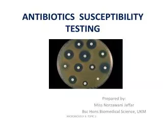
Conventional methods Disk diffusion (Kirby-Bauer) Surface of agar plate seeded with lawn of test organism Inoculum: swab from 0.5 McFarland Disks containing known conc. of agent placed on surface of plate Measure diameter of zone of inhibition
Conventional methods Disk diffusion Zone sizes have been correlated with MICs to establish interpretive criteria Typically, 12 – 13 disks can be placed on each plate
Conventional methods Antibiotic gradient diffusion Agent is applied in gradient to a test strip Plate is seeded with organism as in KB Agent diffuses away from strip to inhibit growth Etest (AB BIODISK, Sweden)
Interpretive categories Susceptible: agent may be appropriate for therapy; resistance is absent or clinically insignificant Intermediate: agent may be useful if conc. at site of infection; may not be as useful as susceptible agent; serves as safety margin for variability in testing Resistant: agent may not be appropriate for therapy; inhibitable dose not acheivable or organism possesses resistance mechanism
Automated systems Manual preparation of isolate suspension Manual – completely automated inoculation Automated incubation, reading of results Automated interpretation and data management
MicroScan WalkAway Dade-Behring