Download
1 / 37
430 likes | 1.18k Vues
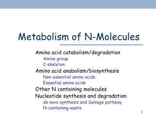
Metabolism of N-Molecules. Amino acid catabolism/degradation Amino group C-skeleton Amino acid anabolism/biosynthesis Non-essential amino acids Essential amino acids Other N containing molecules Nucleotide synthesis and degradation de novo synthesis and Salvage pathway N-containing waste.

E N D
Metabolism of N-Molecules Amino acid catabolism/degradation Amino group C-skeleton Amino acid anabolism/biosynthesis Non-essential amino acids Essential amino acids Other N containing molecules Nucleotide synthesis and degradation de novo synthesis and Salvage pathway N-containing waste
Amino acids catabolism In animals • Protein turnover • Normal cellular protein degradation • ATP-independent process in lysosomes • Ubiquitin-tag + ATP proteasome (p. 1066) • Dietary protein surplus • Amino acids can not be stored • Positive N balance (excess ingestion over excretion) • Growth and pregnancy • Negative N balance (output exceeds intake) • After surgery, advanced cancer, and kwashiorkor or marasmus • Starvation or diabetes mellitus • Protein is used as fuel p. 623
Protein turnover • Membrane associated protein • Lysosome • Cellular protein • Abnormal, damaged, or regulatory proteins. • Ubiquitin (Ub) and proteasome • Ub: the death signal, covalently attached to the target protein • N-terminal rule: (Table 27-10) • Destabilizing residue: Arg, Leu • Stabilizing: Met, Pro • Cyclin destruction boxes • A.a. sequences that mark cell-cycle proteins for destruction • PEST • Proteins rich in Pro, Glu, Ser, and Thr. • Proteasome: executioner • ATP-driven multisubunit protease complex. • Proteasome product: Ub + peptides of 7-9 a.a. • Peptides are further degraded by other cellular proteases. Stryer 5th Fig 23.6
Stryer 5th Stryer 5th Fig 23.3 Biological function • Human papilloma virus (HPV) • Encodes a protein that activates a specific E3 enzyme in ubiquitination process. • E3 Ub the tumor suppressor p53 and other proteins that control DNA repair, when are then destroyed. • E3 activation is observed in 90% of cervical carcinoma. • Inflammatory response • NF-kB (transcription factor) initiates the expression of a number of the genes that take part in this process. • NF-kB normally remains inactivated by binding to an inhibitory protein, I-kB. (NF-kB - I-kB complex) • Signal I-kB phosphorylated I-kB – Ub release NF-kB immune response.
Regulatory enzymes (Review) Fig 8-31 • Polypeptide cleavage : inactive active • Pepsinogen pepsin • Chymotrypsinogen chymotrypsin • Trypsinogen trypsin • Procarboxypeptidase A(B) carboxypeptidase A(B) • Irreversible activation inactivate by inhibitors • Pancreatic trypsin inhibitor (binds and inhibits trypsin) Zymogen or Proprotein or Proenzyme
Stryer 5th Fig 23.1 Protein Digestion • In stomach • Pepsinogen + HCl Pepsin • HCl : denaturing protein exposing peptide bonds • Pepsin cleaves peptide bond before aromatic residues (Table 5-7) • Peptide fragments (7-8 residues) • Pancreas and small intestine • Trypsin (C of Lys, Arg) • Chymotrypsin (C of aromatic a.a.) • Carboxypeptidase, and aminopeptidase free a.a. for absorption • Acute pancreatitis • Obstruction of pancreatic secretion • Premature enzymes attack the pancreatic tissue
Amino acid catabolism • Amino acid = NH3+- + C skeleton • “Bookkeeping” Intracellular protein Dietary protein Amino acids C skeletons NH4+ Citric acid cycle Urea cycle Glucose Fig 18-1 modified CO2 Urea
N-containing wastes (p. 634) p. 625, Fig 18-2(b)
Remove a-amino group • 1st step in liver: transamination • Aminotransferase or transaminase • Exception: proline, hydroxyproline, threonine, and lysine • Collect amino group in glutamate form Fig 18-4 Keto acid Amino acid • Classic example of enzyme catalyzing bimolecular Ping-Pong reactions.
Aminotransferase • A family of enzymes with different specificity for the amino acids. • Alanine aminotransferase • Aspartate aminotransferase • A common prosthetic group (coenzyme): • PLP (pyridoxalphosphate) • Derived from Vit B6 • Transamination • As a carrier of amino group (accept donate) • Decarboxylation • Racimization • Forms enzyme-bound Schiff base intermediate. • Medical diagnoses (Box 18-1) • A variety of enzymes leak from the injured cells into the bloodstream • Heart and liver damages caused by heart attack, drug toxicity, or infection. • Liver damages caused by CCl4, chloroform, and other industrial solvent. • [Enz] in blood serum • SALT test (alanine aminotransferase, or GPT) • SAST test (aspartate …, or GOT) • SCK test (serum creatine kinase)
Mitochondria Cytosol + Urea cycle + + Citric acid cycle Glucose synthesis Glu releases NH4+ in liver • In hepatocytes, Glu is transported from cytosol into the mitochondria. • Glutamate dehydrogenase catalyze the oxidative deamination in mitochondria to release NH4+. • Trans-deamination Fig 18-4 and 18-7
Citric acid cycle Glucose synthesis Urea cycle Glutamate dehydrogenase • Operates at the intersection of N- and C- metabolism • Present only in hepatic mitochondria matrix • Requires NAD+ or NADP+ • Allosterically regulated • Inhibitor: [GTP] and [ATP] • Activator: [GDP] and [ADP] • A lowering of the energy charge accelerates the oxidation of a.a. • Hyperinsulinism-hyperammonemia syndrome: • mutation in GTP binding site, permanently activated. Fig 18-7
a-ketoglutarate + NH4+ Glutamate dehydrogenase NH4+ transport in blood (I) • NH4+ is toxic to animal tissues • Gln is a nontoxic transport form of NH4+ • Gln releases NH4+ in liver and kidney mitochondria by glutaminase In hepatocyte mitochondria In extrahepatic tissues Glu Gln Glutamine synthetase Gln Glu p. 632
Gln TCA cycle (buffer) HCO3- a-ketoglutarate+ NH4+ + acids Salts (excreted) Glutamate dehydrogenase Glu Metabolic acidosis (p. 663) • Kidney extracts little Gln from bloodstream normally • Acidosis increases glutamine processing in kidney • NH4+ + metabolic acids salts (excreted in urine) • a-ketoglutarate bicarbonate (HCO3-, buffer) In kidney kidney’s mitochondria Lehninger 4th ed. Fig 18-8 modified
Fig 18-8 Muscle contraction Gluconeogenesis NH4+ transport in blood (II) • Glucose-alanine cycle • Ala transports NH4+ from skeletal muscle to liver • Pyruvate is recycled to glucose in liver and then returned to muscle • Economy in energy use • Tissue cooperation • Cori cycle (glucose-lactate cycle)
N excretion Most terrestrial animals: • Almost exclusively in liver: • NH4+ urea (urea cycle) • 5 enzymatic steps (4 steps in urea cycle) • 2 cellular compartments involved • Urea bloodstream kidney excreted into urine • Urea cycle and citric acid (TCA) cycle • Regulation of urea cycle • Genetic defect and NH4+ intoxication • Urea cycle defect and protein-rich diet • Essential a.a. must be provided in the diet. • A.A. can not be synthesized by human body. Ch 22 Biosynthesis
Aspartate 3 Arginino-succinate Citrulline 1 2 Carbamoyl phosphate NH4+ + HCO3- 4 Fumarate Arginine 5 Urea (NH2)2CO Urea cycle • Sources of N and C in synthesized (NH2)2CO In the mitochondria and cytoplasm of liver cells • Carbamoly phosphate synthetase I • Ornithine transcarbamoylase • Argininosuccinate synthetase • Argininosuccinate lyase • Arginase Urea Cycle Ornithine Fig 18-9 modified
Sources of NH4+ • Glu and Gln release NH4+ in the mitochondria of hepatocyte • Asp is generated in mitochondrial matrix by transamination and transported into the cytosol of hepatocyte Gln Ala Glu OAA • Refer to Fig 19-26 p. 685 • Malate-Asp shuttle • OAA cannot cross membrane • Malate-aKG transporter • Glu-Asp transporter Asp Fig 18-9 left
Regulation of urea cycle Fig 18-12 p. 636 • Protein-rich diet and prolonged starvation: • urea production. • Long term: • Rate of synthesis of the 4 urea cycle Enz. and carbamoyl phosphate synthetase I in the liver. • Short term: • Allosteric regulation of carbamoyl phosphate synthetase I • Activator: N-acetylglutamate, enhances the affinity of synthetase for ATP.
Carbamoyl phosphate synthetase I • Properties • The 1st enzyme for NH4+ urea • Mitochondria matrix isoform • Type II in cytosol for pyrimidine synthesis (p. 667, and Ch 22) • High conc. than type II in cytosol • Greater need for urea production • Activator: • N-acetylglutamate • acetyl-CoA + Glu • Arginine • Urea cycle defect • N-acetylglutamate synthase deficiency • Supplement with carbomylglutamate (p. 670) Fig 18-13
[NH4+] ↑ [Gln] ↑ H2O uptake ↑ cell swelling [Glu] ↓ [GABA] ↓ [a-KG] ↓ ATP generated from citric acid cycle ↓ NH4+ intoxication (p.665) • Symptoms • Coma • Cerebral edema • Increase cranial pressure • Possible mechanisms • Depletion of ATP in brain cells • Changes of cellular osmotic balance in brain • Depletion of neurotransmitter • Remove excess NH4+ • Glutamate dehydrogenase: NH4+ + a-KG Glu • Glutamine synthetase: NH4+ + Glu Gln
Defect in urea cycle enzymes • Build-up of urea cycle intermediates • Treatments • Strict diet control and supplements of essential a.a. • With the administration of : • Aromatic acids (Fig 18-14) • Lower NH4+ level in blood • Benzoate + Gly + … hippurate (left) • Phenylbutyrate + Glutamine + … phenylacetylglutamine (right) • BCAA derived keto acids • Carbamoyl glutamate (N-acetylglutamate analog) • Deficiency of N-acetylglutamate synthase • Arginine • Deficiency of ornithine transcarbamoylase • Deficiency of argininosuccinate synthetase • Deficiency of argininosuccinase Lehninger 4th ed. p. 669-670
Glucose TCA cycle Energy cost of urea cycle p. 637 • Urea synthesis costs energy… • 4 high energy phosphate groups from 3 ATP • Oxaloacetate (OAA) regenerate produces NADH (Fig 18-11) • 1 NADH 2.5 ATP • Pathway interconnections reduce the energetic cost of urea synthesis • Argininosuccinate shunt Stryer 5th Fig 23.17
Acetone Acetoacetate D-b-hydroxybutyrate Metabolism of C skeleton Amino acid = NH3+- + C skeleton Oxidized to CO2 and H2O Glucose (glucogenic a.a.) Ketone bodies (ketogenic a.a.) Fatty acids oxidation (Ch 17)
Entering citric acid cycle • 20 a.a. enter TCA cycle: • Acetyl-CoA (10) • a-ketoglutarate (5) • Succinyl-CoA (4) • Fumarate (2) • Oxaloacetate (2) • Some a.a. yields more than one end product • Different C fates a-KG TCA cycle Succinyl-CoA Acetyl-CoA Fumarate OAA Fig 18-14
One-carbon transfer p.640-643 • Transfer one-carbon groups in different oxidation states. • Some enzyme cofactors involved (Fig 18-15): • Biotin • Transfer CO2 • Tetrahydrofolate (H4 folate) • Transfer –HC=O, -HCOH, or –CH3 • S-adenosylmethionine (adoMet, SAM) • Transfer –CH3
Threonine Nicotinate (niacin) Serotonin Ala, Trp, Cys, Thr, Ser, Gly Pyruvate Lehninger 4th ed. Fig 18-19 modified
PKU Acetoacetyl-CoA Phe and Tyr Fig 18-21 Top right • Phe + -OH Tyr • Phenylalanine hydroxylase • Phenylketonuria (PKU) • Phe, Tyr as precursor • Fig 22-29, p. 860 • Dopamine • Norepinephrine • Epinephrine • Tyr as precursor • Melanin Phenylalanine hydroxylase
NAD+ H2 biopterin reductase NADH + H+ H4 biopterin Lehninger 4th ed. Fig 18-24 • Phenylalanine hydroxylase • Mixed-function oxidase • Cofactor: tetrahydrobiopterin (H4 biopterin) • Dihydrobiopterin reductase is required to regenerate H4 biopterin • Defect in dihydrobiopterin (H2 biopterin) reductase • PKU, norepinephrine, serotonin, L-dopa deficiency, … • Supplement with H4 biopterin, as well as 5-OH-Trp and L-dopa H4 biopterin H2 biopterin
Branched-chain a.a. (p. 651) • BCAA: Val, Ile, Leu • Not degraded in the liver • Oxidized as fuels in extrahepatic tissues • Muscle, adipose, kidney and brain • The 3 a.a. share the first 2 enzymes for catabolism • Fig 18-27 • Branched-chain aminotransferase a-keto acids • Branched-chain a-keto acid dehydrogenase complex acyl-CoA derivatives • Closely resemble pyruvate dehydrogenase • Inactivated by phosphorylation • Activated by dephosphorylation
Maple Syrup Urine Disease Val, Ile, and Leu (Fig 18-27) Val Ile Branched-chain a-keto acid dehydrogenase complex Branched-chain Aminotransferase Leu a-keto acids
Maple syrup urine disease p. 652 • MSUD • Branched-chain ketonuria • Defective branched-chain a-keto acid dehydrogenase complex • a-keto acids (odor) derived (Val, Ile and Leu) accumulate in blood and urine • Abnormal brain development • Mental retardation • Death in infancy • Rigid diet control • Limit the intake of Val, Ile, Leu to min. requirement for normal growth
Genetic disorders • Caused by defective catabolic enzymes
Ketogenic vs. glucogenic a.a. • Acetyl-CoA • Ketone bodies • OAA • a-ketoglutarate • Succinyl-CoA • Fumarate • Gluconeogenesis Acetyl-CoA OAA • Ketogenesis • Glucogenesis Fig 18-29
Ketogenesis vs. glucogenesis • Ketogenesis • A.A. degraded to acetoacetyl-CoA and or acetyl-CoA (6 a.a.) • Yield ketone bodies in the liver • In untreated diabetes mellitus, liver produces large amounts of ketone bodies from both fatty acids and the ketogenic a.a. • Exclusively ketogenic: Leu and Lys • Glucogenesis • A.A. degraded to pyruvate, a-ketoglutarate, succinyl-CoA, fumarate, and/or oxaloacetate • Converted into glucose and glycogen. • Both ketogenic and glucogenic • Phe, Tyr, Trp, and Ile On p. 588, read the 1st paragraph under “The Glyoxylate Cycle”
Biosynthesis C-skeleton NH4+ Shunt Citric acid cycle Urea cycle Excretion Gluconeogenesis Catabolism of a.a. in mammals Fig 18-1, 18-11 modified • The NH3+ and the C skeleton take separate but interconnected pathways Amino acids Fumarate Malate AspOAA
Vit B12 and folate (p. 674) • Met synthesis in mammal • N5-methyl H4 folate as C donor • C is then transferred to Vit B12 • Vit B12 as the final C donor • Vit B12 deficiency • H4 folate is trapped in N5-methyl form (formed irreversibly) • Available folate ↓ • e.g. pernicious anemia Lehninger 4th ed. Fig 18-18 left