
Uploaded by
tynice
1 SLIDES
116 VUES
10LIKES
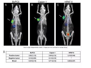
Analysis of .BxPC3, Capan-1, and HPAF-II Cell Line Images with Independent Scaling
DESCRIPTION
This study presents images derived from the pancreatic cancer cell lines .BxPC3, Capan-1, and HPAF-II. Each image has been independently scaled, providing a unique perspective on cellular morphology and features. It is important to note that the images have not been corrected for isotope decay, which may affect the accuracy of quantified results. This analysis aims to assist researchers in understanding the characteristics of these cell lines for further studies in cancer research.
Download
1 / 1
Télécharger la présentation 
Analysis of .BxPC3, Capan-1, and HPAF-II Cell Line Images with Independent Scaling
An Image/Link below is provided (as is) to download presentation
Download Policy: Content on the Website is provided to you AS IS for your information and personal use and may not be sold / licensed / shared on other websites without getting consent from its author.
Content is provided to you AS IS for your information and personal use only.
Download presentation by click this link.
While downloading, if for some reason you are not able to download a presentation, the publisher may have deleted the file from their server.
During download, if you can't get a presentation, the file might be deleted by the publisher.
E N D
Presentation Transcript
BxPC3 Capan-1 HPAF-II A B C Each image independently scaled. Images are not corrected for isotope decay D


More Related