Download
1 / 43
520 likes | 1.43k Vues
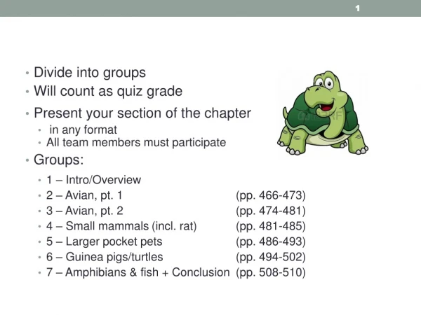
Divide into groups Will count as quiz grade Present your section of the chapter in any format All team members must participate Groups: 1 – Intro/Overview 2 – Avian, pt. 1 (pp. 466-473) 3 – Avian, pt. 2 (pp. 474-481) 4 – Small mammals (incl. rat) (pp. 481-485)

E N D
Divide into groups • Will count as quiz grade • Present your section of the chapter • in any format • All team members must participate • Groups: • 1 – Intro/Overview • 2 – Avian, pt. 1 (pp. 466-473) • 3 – Avian, pt. 2 (pp. 474-481) • 4 – Small mammals (incl. rat) (pp. 481-485) • 5 – Larger pocket pets (pp. 486-493) • 6 – Guinea pigs/turtles (pp. 494-502) • 7 – Amphibians & fish + Conclusion (pp. 508-510)
Dental Radiography Lavin: Chapter 24 "Do not be daunted by the enormity of the world's grief. Do justly, now. Love mercy, now. Walk humbly, now. You are not obligated to complete the work, but neither are you free to abandon it." - The Talmud
Objectives: Dental Radiography • Understand dental terminology & tooth surfaces • Understand basic anatomy & formula for teeth, including number of roots • Understand normal views & positioning for dental radiography • Define parallel and bisecting angle techniques, and know when to use each • Understand the differences between intraoral and extraoral views
Dental Radiography: Overview • Includes both intraoral and extraoral radiographs • Special equipment isn’t essential • Allows thorough evaluation of tooth & surrounding tissues • Indications for dental radiographs: • Periodontal disease • Missing, malformed, or discolored teeth • Resorptive lesions • Oral tumors • Gingival inflammation • Trauma • Dental extractions
Positional Terminology: Oral Cavity Directional Positional
Tooth Anatomy: A Review • 4 tooth types: • Incisors – used for grooming, grasping, cutting • Canine teeth – grasping/holding prey • Premolars – cutting/shearing • Molars – grinding (dogs), cutting (cats)
Dental Formulas • Carnassial tooth: • Shearing cheek tooth • Dogs: Upper 4th premolar & lower 1st molar • Cats: Upper 3rd premolar & lower 1st molar
Triaden Numbering System Canine Feline Canine Feline • 1st number = quadrant • 2nd 2 numbers = tooth position
Normal Adult Canine Maxillary Incisors 7 – Incisive canal 8 – Interincisive suture 9 – Palatine fissure – oval dark spaces
Normal Adult Canine Mandibular Incisors 7 – Mandibular symphysis
Normal Adult Canine Maxillary Premolars/Molars 8 – Nasal cavity 11 – Palatal canal
Normal Adult Canine Mandibular Premolars/Molars 9 – Mandibular canal 11 – Enamel overlap
Viewing Dental Radiographs • Film is exposed with the convex dot at the rostral end of the mouth • Dot location varies with right & left radiographs
Film Mount Organize full mouth radiographs in a film mount: Hang as if looking at the animal, with animal’s right side on your left…
Viewing Dental Radiographs (Cont.) • After film is developed, hold it as if in the mouth with the raised dot towards you • Visualize the film inside the animal’s mouth • Determine via anatomy whether mandible or maxilla • Cusps of maxillary teeth should point down • Mandibular teeth point up
Radiography Techniques Parallel Bisecting Angle
Dental: Parallel Technique • Used for mandibular molars and pre-molars only • Film is placed inside of & parallel to the teeth • Primary bean is placed perpendicular to film
Dental: Bisecting-angle Technique • For all maxillary teeth & mandibular incisors • Placing the beam perpendicular to tooth or film results in foreshortening or elongation, so bisecting-angle minimizes • Find the bisecting-angle: • Place film parallel to tongue • Draw an imaginary line along the tooth’s axis • Draw another imaginary line along the axis of the film • Position the primary beam perpendicular to the “bisecting angle”
Bisecting Angle CTVT pg. 888
Types of Dental Radiography Digital • Dental Film-Based X-ray unit • Digital Radiography • Regular X-ray machine • Intraoral – Film inside the mouth • Extraoral – Film outside the mouth Regular Machine - Intraoral Regular Machine - Extraoral
Dental Radiography Can be done in two ways: • Conventional machine • Not ideal, but may be only option • Patient’s body position/skull must be rotated • Positioning very difficult • Screened cassette & radiolucent devices required • Distal teeth may be difficult to image • SID (source to image distance) must be reduced to 15 inches or exposure factor adjustments must be made • Dental X-Ray machine • Smaller & portable • Radiographs of superior quality • Expensive Feline
Conventional Dental Radiography: Considerations • Not ideal, but may be all that’s available • Requires screened cassettes & radiolucent positioning devices • Positioning the cassette in the mouth is usually difficult • Distal teeth are especially difficult to image • Bone superimposition can make molars & premolars appear whiter
Conventional Radiology: Extraoral Maxillary Premolars/molars: VD Oblique Extraoral View • Patient positioned halfway between lateral & VD • Affected side down • Place foam wedge/roll under nose • Rotate mandible to 45-degree angle from table surface • Place radiolucent mouth gag
Conventional Radiology: Intraoral Maxillary Incisors & Canine: DV Intraoral/Occlusal View • Sternal recumbency • Place sponge under mandible & cassette to keep parallel to table • One corner of cassette into patient’s mouth • Adjust SID for raised cassette • Can assess rostral nasal sinus cavity
Dental Radiography: Dental Machine • Size of film depends on animal & tooth • Lateral patient positioning w/jaws parallel to tabletop • Use foam or wedge as needed • Tie ET to opposite jaw from area of interest • Paper towel can be used to position film in mouth • SID varies from against face (small film) to 6 inches • Increased SID = increased exposure • No collimator, so estimate • Place dot rostrally
Radiography: Maxillary Canine Tooth • Initially set up using the long axis of the tooth • Cone is directed obliquely toward animal’s midline • Prevents superimposition of canine tooth over premolars • Use root of tooth for bisecting angle, not crown • Both canine teeth should touch film • SID usually 6 inches
Radiography: Mandibular Incisors • Rostrocaudal view can capture all incisors on one film • Roots will overlap slightly • Elongation is common
Radiography: Feline Maxillary Premolars • Intraoral technique • Tongue should be behind the film on the mandible • ET tube will be on the tube side of the film • May have to be untied and clamped to fur on one side • Uses modified parallel technique with beam perpendicular to film
Error: Root Cutoff Not pushing film in far enough
Error: Cone Cutoff Cone not centered on tooth
Artifact: Light Exposure Light exposure in chairside darkroom
Error: Film Processing Fixer not rinsed off film
Error: Bent Film Crescent Line