Pedigrees and Karyotypes
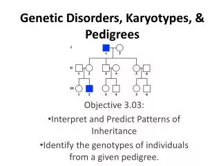
Pedigrees and Karyotypes. Pedigree. A pedigree shows the relationships within a family and it helps to chart how one gene can be passed on from generation to generation.


Pedigrees and Karyotypes
E N D
Presentation Transcript
Pedigree • A pedigree shows the relationships within a family and it helps to chart how one gene can be passed on from generation to generation. • Pedigrees are tools used by genetic researchers or counselors to identify a genetic condition running through a family, they aid in making a diagnosis, and aid in determining who in the family is at risk for genetic conditions.
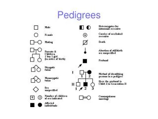
On a pedigree: • A circle represents a female • A square represents a male • A horizontal line connecting a male and female represents a marriage • A vertical line and a bracket connect the parents to their children • A circle/square that is shaded means the person HAS the trait. • A circle/square that is not shaded means the person does not have the trait. • Children are placed from oldest to youngest. • A key is given to explain what the trait is.
Marriage Has the trait Female-daughter Female-daughter Male-Son Male- Son Oldest to youngest Male-DAD Female-MOM
ff ff Ff ff Ff Key: affected male affected female unaffected male unaffected female Ff • Steps: • Identify all people who have the trait. • For the purpose of this class all traits will be given to you. In other instances, you would have to determine whether or not the trait is autosomal dominant, autosomal recessive, or sex-linked. • In this example, all those who have the trait are homozygous recessive. • Can you correctly identify all genotypes of this family? • F- Normal • f- cystic fibrosis
Key: affected male affected female unaffected male unaffected female Pp Pp • PKU • P- Unaffected • p- phenylketonuria Pp pp PP or Pp pp pp Pp Pp
Key: affected male affected female unaffected male unaffected female hh Hh • H-huntington’s disease • h-Unaffected Hh hh Hh hh hh Hh hh
Key: affected male affected female unaffected male unaffected female Sex-Linked Inheritance Cy cc • Colorblindness cy Cc Cc cy cy
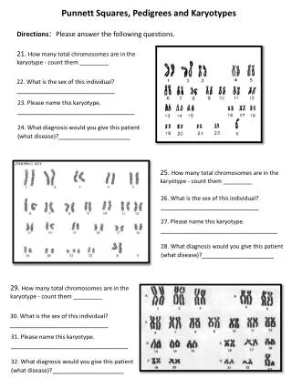
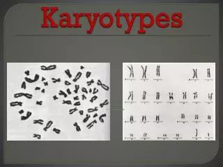
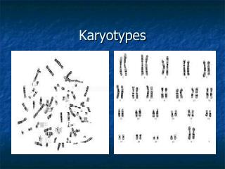
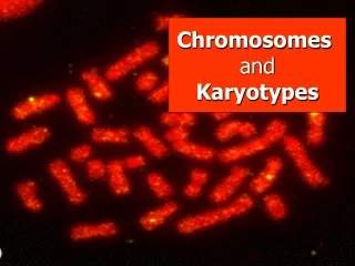
Karyotypes • To analyze chromosomes, cell biologists photograph cells in mitosis, when the chromosomes are fully condensed and easy to see (usually in metaphase). • The chromosomes are then arranged in homologous pairs.
Karyotypes • The homologous pairs are then placed in order of descending size. The sex chromosomes are placed at the end. • A picture of chromosomes arranged in this way is known as a karyotype.
Karyotypes • The karyotype is a result of a haploid sperm (23 chromosomes) fertilizing a haploid egg (23 chromosomes). • The diploid zygote (fertilized egg) contains the full 46 chromosomes. (in humans)
Labeling a Karyotype • To label a karyotype correctly, first list the number of chromosomes found in the karyotype. Ex. 46 • Secondly, list the type of sex chromosomes found in the karyotype. Ex. XX • Lastly, list the any abnormalities at the appropriate chromosome number. Normal Human Female: 46, XX Normal Human Male: 46, XY
What are abnormalities? • Sometimes, during meiosis, things go wrong. • The most common error is nondisjunction, which means “not coming apart”. • If nondisjunction occurs , abnormal numbers of chromosomes may find their way into gametes, and a disorder of chromosome numbers may result.
Autosomal Chromosome Disorders • Two copies of an autosomal chromosome fail to separate during meiosis, an individual may be born with THREE copies of a chromosome. • This is known as a “Trisomy” • Trisomy 13, Trisomy 18, Trisomy 21.
Male: 47, XY, +21 Female: 47, XX, +21 Down Syndrome • Most common, Trisomy 21 (down syndrome) • 1 in 800 babies born in U.S. with Trisomy 21. • Mild to severe mental retardation • Increased susceptibility to many diseases and a higher frequency of other birth defects.
Klinefelter’s Syndrome, 47 XXY Sex Chromosome Disorders • Turner’s Syndrome (nondisjunction) • Female inherits only one X chromosome • Karyotype: 45, X • Women are sterile, sex organs do not develop at puberty. • Klinefelter’s syndrome (nondisjunction) • Males receive an extra X chromosome • Karyotype: 47, XXY • The extra X chromosome interferes with meiosis and prevents ind. from reproducing.
Other Genetic Disorders • Sickle Cell Disease • Characterized by the bent and twisted shape of the red blood cells. • More rigid and get stuck in capillaries. Blood stops flowing and can damage cells, tissues, and organs. • Produced physical weakness and damage to the brain, heart, and spleen…could be fatal. • Most commonly found in African Americans (can be linked to the incidence of malaria).
Other Genetic Disorders • Duchenne Musclular Dystrophy • Sex-linked, defective gene for muscle protein. • Progressive weakening and loss of skeletal muscle. • In U.S., 1 out of every 3000 males born has condition.
On the schedule! • Karyotype Lab • Genetic Disorder Project and Presentations • . SPRING BREAK!!!