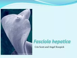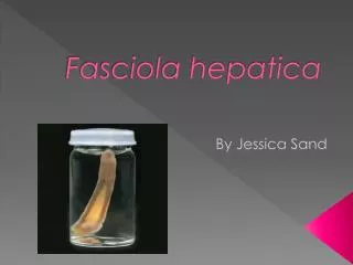
Fasciola hepatica
Fasciola hepatica. Lab Miss Hiba Bourinee. Medically significant trematodes. Fasciola hepatica. Fasciola hepatica , also known as the common liver fluke or sheep liver fluke .

Fasciola hepatica
E N D
Presentation Transcript
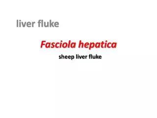
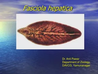
Fasciola hepatica Lab Miss Hiba Bourinee
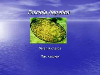
Fasciola hepatica • Fasciola hepatica, also known as the common liver fluke or sheep liver fluke. • Is a parasitic flatworm of the class Trematoda, phylum Platyhelminthes that infects liver of various mammals, including humans. • The disease caused by the fluke is called fascioliasis (also known as fasciolosis). • F. hepatica is world-wide distributed and causes great economic losses in sheep and cattle.
Life cycle • In order to complete its life cycle, F. hepatica requires an aquatic snail as an intermediate host such as Galba truncatula, in which the parasite can reproduce asexually. • From the snail, minute cercariae emerge and swim through pools of water in pasture, and encyst as metacercariae on nearby vegetation.
Life cycle • From here, the metacercariae are ingested by the ruminant, or in some cases, by humans eating un-cooked foods such as water-cress. • Contact with low pH in the stomach causes the early immature juvenile to begin the process of excysment . • In the duodenum, the parasite breaks free of the metacercariae and burrows through the intestinal lining into the peritoneal cavity.
Life cycle • The newly excysted juvenile does not feed at this stage, but once it finds the liver parenchyma after a period of days, feeding will start. • This immature stage in the liver tissue is the pathogenic stage, causing anaemia and clinical signs sometimes observed in infected animals.
Life cycle • The parasite browses on liver tissue for a period of up to 5-6 weeks and eventually finds its way to the bile duct where it matures into an adult and begins to produce eggs. • Up to 25,000 eggs per day per fluke can be produced, and in a light infection, up to 500,000 eggs per day can be deposited onto pasture by a single sheep.
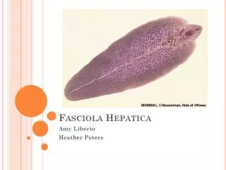
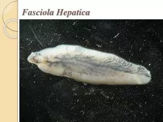
Morphology • Adult worms are large leaf-like, 20 to 30 mm in length. • At the anterior end, distinct conical projection is observed. • The ceca and testes are highly branched. • The eggs measure about 140 by 80 µm.
egg capsule with emerging miracidium of Fasciola hepatica. 400x
Pathology and clinical symptoms. • The triad of fever, hepatomegaly, and eosinophilia in endemic area suggests fascioliasis. • Symptoms and signs are associated with biliary obstruction and cholangitis. • Acute epigastric pain, pruritus, and jaundice are common. • Worms in human infections may be frequently found in ectopic foci.
Prevention treatment • Avoiding ingestion of raw watercress grown in endemic areas.
Treatment of F.hepatica • The drug of choice in the treatment of fasciolosis is triclabendazole, a member of the benzimidazole family of anthelmintics. • However, resistance of F. hepatica to triclabendazole has already been recorded in Australia and Ireland. • Artemether has been shown to be effective in a rat model of fascioliasis .
Fasciolopsis buski • Fasciolopsiasis results from infection by the trematode Fasciolopsis buski, the largest intestinal fluke of humans (up to 7.5 cm in length) Geographic distribution • This disease occurs in Asia and the Indian subcontinent, especially in areas where humans raise pigs and consume raw aquatic plants.
Clinical features • Most infections are light and asymptomatic. In heavier infections, symptoms include diarrhea, abdominal pain, fever, ascites, anasarca, and intestinal obstruction.
F.buski morphology • Egg are yellow brownoval large 75-100µm x 130-150µm, and surrounded by a thin shell. • They are unempryonated when passed.these eggs are identical to those of F.hepatica,and when seen should be reported as F.buski/F.hepatica eggs. • Large adult flukes 1.5 cm x 3 cm2-7 cm long lack the cephalic cone seen in F.hepatica.
Life cycle • Though many of us are familiar with the term “liver fluke,” F. buski does not live in the liver: these large leaf-shaped worms inhabit the upper regions of the small intestine. • When there are many worms, they are also found in lower areas of the intestine and in the stomach. Fasciolopsis buski is not found in other parts of the body
This photo is to compare the sizes of Fasciolopsis buski (left) and Fasciola hepatica (right), 2x.
F.buski • Immature unembryonated eggs are discharged into the intestine and stool . • Each egg becomes embryonated (3-7 weeks) in warm water. • When fully developed, egg hatchtes to release miracidium. • Miracidium invades a suitable planorbid snail intermediate host. In the snail the parasites undergo asexual development (generations of sporocysts, redia then cercaria ).
F.buski • Cercaria are released from the snail. • Cercaria then encyst as metacercaria on aquatic plants particularly Trapa natans (the water caltrop) or the water chestnut, which thrives in ponds fertilised by "night soil" (human faeces) . • The mammalian hosts become infected by ingesting metacercariae on the aquatic plants.
F.buski • Note: most common mammalian hosts are pigs, humans and dogs. • Note: infection occurs if the person uses his teeth to peel the outer covering of the infected plants (the inside, the only edible part, is free of worms).
F.buski life cycle • After ingestion, the metacercariae excyst in the duodenum and attach to the mucosa of the jejunal and duodenal wall (intestinal wall). • There they develop into adult flukes (20 to 75 mm by 8 to 20 mm) in approximately 3 months, attached to the intestinal wall of the mammalian hosts (humans and pigs). • the largest intestinal fluke of humans (up to 7.5 cm in length).
F.buski life cycle buski • The adults have a life span of about 6 months (some sources say up to one year). • They go through sexual reproduction to make the opperculated eggs that are then dischared into the intestine and exit in stool.
F.buski diagnosis • Microscopic identification of eggs, or more rarely of the adult flukes, in the stool or vomitus is the basis of specific diagnosis. The eggs are indistinguishable from those of Fasciola hepatica.
F.buski diagnosis • Enzyme-linked immunosorbent assay (ELISA) are also used for diagnosis of ectopic fascioliasis. • Radiologic techniques can provide indirect evidences of fascioliasis.
Treatment of F.buski • The drug of choice is praziquantal, and the alternative is niclosamide. Prevention • Proper sanitation, • control of human feces • Ellimination of snail population.