Rotifera
“ wheel Animalcules ”. Rotifera. Rotifers were first described in 1969 by John Harris. There have been descriptions of about 2200 species of Rotifers. The four classes of Rotifera are the Monogononta , the Bdelloidea , the Seisonidea , and the Digononta .


Rotifera
E N D
Presentation Transcript
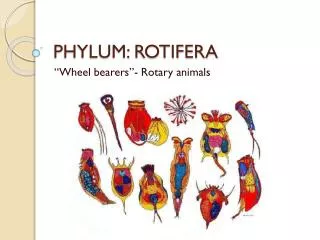
“wheel Animalcules” Rotifera
Rotifers were first described in 1969 by John Harris. There have been descriptions of about 2200 species of Rotifers. The four classes of Rotifera are the Monogononta, the Bdelloidea, the Seisonidea, and the Digononta. The word rotifer is derived from a Latin word that means “wheel-bearer”. This is because the corona around the mouth of a rotifer would resemble a wheel if in motion. The class Monogononta has the most species (1500), followed by the Bdelloidea (350). History of Rotifers
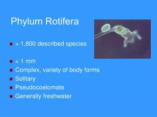
Rotifers are microscopic creatures of the phylum rotifera. Rotifers can live in still water environments such as lakes or flowing water environments such as rivers or streams. Rotifers are commonly found on mosses, lichens, and mushrooms. Rotifers are not commonly fossilized due to the fact that they have soft bodies. A rotifers jaw is its only hard part. Rotifers are recognized as animals even though they are microscopic because they have specialized organ systems and a complete digestive tract. Rotifers
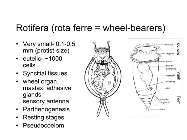
The general rotifer’s body consists of four different regions. The head, the neck, the trunk, and the foot. The head has a corona of cilia that sucks water into the mouth. The rotifer sifts through the water for food. The food is ground up by the trophi (jaws) that are located behind the mouth in the throat. Trophi are characteristic organs of the phylum rotifera. Within the trunk are the stomach and the reproductive organs. The toe contains a cement gland. Physical Characteristics of Rotifers
Philodina are some of the most frequently encountered rotifers. Philodina are large and transparent. Their organs are easily observed at high magnification. Philodina have two ovaries. To feed, Philodina attach themselves to objects and sift through the water for smaller organisms and particles of debris. Females only produce females. Philodina
Brachionus are found in freshwater and marine habitats. Brachionus have transparent, turtle like shells. Some Brachionus are cultured to provide food for fish larvae. Brachionus carry their eggs on their tales. Brachionus
Rotifer vulgaris is frequently encountered in moss. Polyarthravulgaris has 4 blade like projections or paddles. These paddles stream behind polyarthravulgaris as it swims. Common predators of the polyarthravulgaris include protozoans, cladocerans, and copepods. PolyarthraVulgaris
Lecane have shells and live in freshwater bodies. The dorsal plates of Lecane are covered with ridges and folds. These ridges are important to identifying its species. Lecane
Collotheca does not possess a corona of cilia but rather a net of tentacles to bring in food. Collotheca do not move often. A Collotheca carries its egg on its foot. When they do move, they attach to each other to form a spherical colony. Collotheca
http://en.wikipedia.org/wiki/Rotifer http://www.ucmp.berkeley.edu/phyla/rotifera/rotifera.html http://www.microscopy-uk.org.uk/mag/artmar04/jmcrotif.html http://www.micrographia.com/specbiol/rotife/homebdel/bdel01ph.htm http://micro.magnet.fsu.edu/moviegallery/pondscum.html http://www.microscopy-uk.org.uk/mag/wimsmall/extra/rotif7.html Sources