MODULE 3 CHAPTER 1D
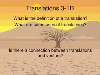
MODULE 3 CHAPTER 1D. VASCULAR AGING. PLAN. 1. Introduction 2. Normal vascular anatomy and physiology 3. What is vascular aging ? 4. What causes it ? 5. How to diagnose it ? 6. How to treat and prevent it ? 7. Take away. Aging : What matters?. 45 yr IT professional

MODULE 3 CHAPTER 1D
E N D
Presentation Transcript
PLAN 1. Introduction 2. Normal vascular anatomy and physiology 3. What is vascular aging ? 4. What causes it ? 5. How to diagnose it ? 6. How to treat and prevent it ? 7. Take away
Aging : What matters? 45 yr IT professional Arteries are 70 yr old 70 yr Athlete Arteries are 45 yr old Chronological age is different from biological age
Atherothrombosis Significantly Shortens Life Atherothrombosis reduces life expectancy by around 8-12 years in patients aged over 60 years1 Average Remaining Life Expectancy at Age 60 (Men) 20 -7.4 years 16 -9.2 years -12 years 12 Years 8 4 0 Healthy History of Cardiovascular Disease History of AMI History of Stroke Analysis of data from the Framingham Heart Study. Peeters A, et al. Eur Heart J. 2002;23:458-466.
Vascular age • Vascular age is the apparent age of the blood vessels, particularly the arteries when compared to what is normal for the healthy population • Vascular age is affected by genetic predisposition, lifestyle choices and other factors
PLAN 1. Introduction 2. Normal vascular anatomy and physiology 3. What is vascular aging? 4. What causes it? 5. How to diagnose it? 6. How to treat and prevent it? 7. Takeaways
THE ARTERIAL SYSTEM • These are the main conducting vessels carrying blood away from the heart • Can be muscular or elastic. There is a gradual transition between these two types
Elastic Arteries • Are characterised by a predominance of elastin in the tunica media and little smooth muscle • Are found just downstream from the heart • Undergo expansion with each systole of the heart • On relaxation of the heart the elastic recoil of the wall helps propel blood through the blood vessels • Include the aorta, pulmonary, common carotid and other major vessels
Muscular Arteries • These are medium sized to smaller arteries • Are characterised by a predominance of smooth muscle cells in the tunica media • Make up the main distributing branches of the arterial tree, eg. the femoral, radial, coronary and cerebral arteries • Remember, there is a gradual transition between elastic and muscular arteries.
ARTERIES ARTERIOLES AORTA ELASTIC MUSCULAR
Increases vascular afterload with a propensity to develop LVH Decreases coronary perfusion pressure Increases myocardial oxygen demand and subendocardial ischemia Increases flow turbulence, endothelial dysfunction and atherogenesis Increases in pulsatile strain and chance of plaque rupture All recognized by a wide brachial artery pulse pressure in the elderly Elderly stiff arteries with ISH : Increased PW velocity (12 m/sec) Normal Arterial Pulsation Young compliant arteries :Normal PW velocity (8 m/sec) Diastole Systole Cushion Conduit (1) Ventricular-Vascular coupling (2) coronary blood flow Forward wave Systole Reflected Wave (1) Ventricular-vascular mismatch (2) The reflected wave increases or “augments” central SBP during late systole:
Conduit function – Role of Endothelium Tunica adventitia Tunica media Tunica intima Endothelium Subendothelial connective tissue Internal elastic membrane Smooth muscle cells Elastic/collagen fibers External elastic membrane
Normal Endothelium The endothelium is the gatekeeper of the vasculature and a major regulator of vascular tone and hemostasis It Provides a smooth, non-thrombogenic surface and a selectively permeability barrier between the circulation and the vessel wall. Regulates vascular tone and produces • Endogenous vasodilators and vasoconstrictors. • Growth promoting and growth inhibiting factors. • Anticoagulants and Procoagulants. Retards platelet and leucocyte adhesion Inhibits VSMC migration & proliferation Barrier to LDL, degrade VLDL & chylotriglyceride Adapted from Ormolgul and Dzau, J Vasc Med Biol., 1991, 282-301, Pepine, C., et. al., “Vascular Health as as Therapeutic Target in Cardiovascular Disease,” VBW , University of Florida, 1998 Atomic force micrograph frpm Barbara et al, Am J Physiol [Heart Care Physiol.], 1995; 266: H1765-H1772
Normal Arterial Function • Cushion function – mainly by Aorta; prevents transmission of systolic pressure to the periphery , slows systolic velocity and maintains continuous flow in distributing arteries and arterioles • Conduit function – Mainly by arteries and their endothelium ; prevents atheroma, plaque, thrombus formation to provide continuous and uninterrupted blood flow to the organs
PLAN 1.Introduction 2.Normal vascular anatomy and physiology 3.What is the mechanism of vascular aging ? 4.What causes it ? 5.How to diagnose it ? 6.How to treat and prevent it ? 7.Takeaways
Mechanism • Loss or reduction of Cushion and Conduit functions of the blood vessels
YOUNG OLD NORMAL SIZE NORMAL RELAXATION DILATED STIFF
Elderly stiff arteries with ISH : Increased PW velocity (12 m/sec) Aortic Stiffening and Early Wave Reflection Young compliant arteries :Normal PW velocity (8 m/sec) Systole Diastole (1) Ventricular-Vascular coupling (2) coronary blood flow Systole (1) Ventricular-vascular mismatch (2) The reflected wave increases or “augments” central SBP during late systole:
Abnormal cushion function LV systolic,diastolic dysfunction
Abnormal Conduit FunctionAbnormal Endothelium LDL-C Hypertension Angiotensin II Homocysteine Diabetes Smoking Endothelial dysfunction sets the stage for atherosclerosis Oxidative stress Oestrogen deficiency Dysfunction Formation of superoxide anion with inactivation of NO & stimulation of vascular oxidase system platelet and leucocyte adhesion VSMC migration & proliferation LDL deposition lipid clearance Adapted from Ormolgul and Dzau, J Vasc Med Biol., 1991, 282-301 Griendling, K. et.al., “Oxidative Stress and Cardiovascular Disease,”Circulation, 1007; 96: 3264-3265. Atomic force micrograph frpm Barbara et al, Am J Physiol [Heart Care Physiol.], 1995; 266: H1765-H1772
Foam Cells Fatty Streak Fibrous Plaque Complicated Lesion/Rupture Intermediate Lesion Atheroma Endothelial Dysfunction The Evolution of Atherosclerosis From 1st Decade From 3rd Decade From 4th Decade Growth Mainly by Lipid Accumulation Smooth Muscle & Collagen Thrombosis, Hematoma Adapted From Stary HC et al. Circulation. 1995;92:1355-1374
Abnormal conduit function Abnormal cushion function VASCULAR AGING
PLAN 1. Introduction 2. Normal vascular anatomy and physiology 3. What is the mechanism of vascular aging ? 4. What causes it ? 5. How to diagnose it ? 6. How to treat and prevent it ? 7. Takeaways
INTERHEART: the Effect of Modifiable Factors on Risk for MI FAMILY HISTORY Midsegment obesity
LARGE ARTERIES HYPERGLYCEMIA,OTHER RF A G E S PROTEIN GLYCATION IN VESSEL WALL LESS DISTENSIBLE COLLAGEN ATHEROMA HIGH SYSTOLIC BP LOSS OF ARTERIAL COMPLIANCE LOW DIASTOLIC BP HIGH PULSE PRESSURE
Factors contributing to Abnormal conduit function LDL-C Hypertension Angiotensin II Homocysteine Diabetes Smoking Oxidative stress Endothelial dysfunction NO + Local mediators + Tissue ACE, Angiotensin II PAI-1 VCAM, ICAM, Cytokines- NF-kB Endothelin Growth factors, matrix Proteolysis Thrombosis Inflammation Vasoconstriction Vascular lesion and remodelling Plaque rupture Clinical Sequelae Gibbons GH, Dzau VJ, New England J Med,1994; 330: 1431-1438
Focal, Occlusive Intimal disease Inflammatory Endothelial dysfunction Related to LDL cholesterol oxidation “Inside-out” Sensitive to A II and other substances Diffuse, Dilatory Medial disease Fibrotic (elastin breakdown, collagen increase) Adventitial and medial hypertrophy Related to age and BP “Outside-in” Sensitive to A II and other substances ATHERO- ARTERIO-SCLEROSIS SCLEROSIS (Increased vascular stiffness Decreased vascular compliance)
PLAN 1.Introduction 2.Normal vascular anatomy and physiology 3.What is the mechanism of vascular aging ? 4.What causes it ? 5.How to diagnose it ? 6.How to treat and prevent it ? 7.Takeaways
(CUSHION) (CONDUIT)
Identification of Arterial Aging ABNORMAL CUSHION EFFECT NORMAL CUSHION EFFECT High systolic,low Diastolic and high Pulse pressure Normal systolic,diastolic And pulse pressure
CAD Death Rate per 10,000 Person-years 80.6 48.3 43.8 38.1 37.4 34.7 31.0 25.3 25.8 25.2 24.9 24.6 23.8 160+ 16.9 13.9 12.6 12.8 11.8 20.6 140-159 10.3 11.8 8.8 8.5 9.2 120-139 Systolic BP (mmHg) <120 100+ 90-99 80-89 75-79 70-74 <70 Diastolic BP (mmHg) Neaton et al. Arch Intern Med 1992; 152:56-64. Effect of SBP and DBP onAge-Adjusted CAD Mortality: MRFIT High systolic and low diastolic pressure is dangerous
Blood pressure in the aorta, closer to the vital organs What is Central Aortic Pressure ? Central aortic pressure Peripheral brachial pressure
RISK FACTORS PREDICT DISEASE DM,DUR. LIPIDS HBP SMOKING MET SYN MENTAL STRESS FAMILY HISTORY RISK MARKERS INDICATE PRESENCE MICROALB.,ED,CKD, FMD CIMT, AB INDEX,PW VELOCITY MSCT, STRESS TEST Hs CRP ECHO – DD,E/E’ RISK FACTORS AND MARKERS
INDIVIDUAL ASSESSMENT FOR EARLY VASCULAR AGING (EVA)? AT 30 YRS RISK FACTOR AND RISK MARKER ASSESSMENT RISK FACTORS + RISK MARKERS + RISK FACTORS – RISK MARKERS -- RISK FACTORS + RISK MARKERS -- ALREADY EVA + LIKELY TO DEVELOP EVA PREVENT EVA
PLAN 1.Introduction 2.Normal vascular anatomy and physiology 3.What is the mechanism of vascular aging ? 4.What causes it ? 5.How to diagnose it ? 6.How to treat and prevent it ? 7.Takeaways
To reverse and prevent vascular aging • Life style modification – Diet/ Exercise / Good habits / Mental relaxation • Block Renin- Angiotensin system • Control Blood pressure • Reduce Lipids • Control Blood sugar
Local Angiotensin System in Macrophages and Role in Atherosclerosis LDL lumen ACE circulating monocytes ACE Angiotensin II + + Y Y Y Y Y Y Y Y adhesion- molecules infiltration endothelium + oxidative stress endothelial damage ACE oxLDL/eLDL subintima differentiation (activation) fatty streak, plaque ACE ACE Macrophages Foam-cells + smooth muscle cells Growth factors Ang II Ang II Cytokines media Mod. from Diaz et al., N Engl J Med 337 (1997)
ACE I OR ARB? WHICH ACEI,ARB?
TELMISARTAN LIFE Charm