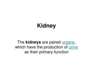
The kidney
This resource provides an in-depth examination of kidney assessment, including the definition of excretion and osmoregulation. It features detailed diagrams of the kidney and glomerulus, highlighting their functional components. The text explains ultrafiltration, focusing on blood pressure, fenestrated capillaries, and the basement membrane. It also covers the reabsorption processes in the proximal convoluted tubule, highlighting the roles of microvilli, osmosis, and active transport. Further discussions include the Loop of Henle, the action of ADH, and variations in the concentration of substances in blood plasma, glomerular filtrate, and urine.

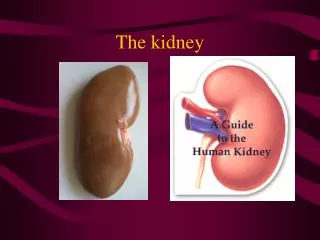
The kidney
E N D
Presentation Transcript
The kidney Topic 11.3
Assessment Statements 11.3.1 Define excretion. 11.3.2 Draw and label a diagram of the kidney. 11.3.3 Annotate a diagram of a glomerulus and associated nephron to show the function of each part. 11.3.4 Explain the process of ultrafiltration, including blood pressure, fenestrated blood capillaries and basement membrane. 11.3.5 Define osmoregulation. 11.3.6 Explain the reabsorption of glucose, water and salts in the proximal convoluted tubule, including the roles of microvilli, osmosis and active transport. 11.3.7 Explain the roles of the loop of Henle, medulla, collecting duct and ADH (vasopressin) in maintaining the water balance of the blood. 11.3.8 Explain the differences in the concentration of proteins, glucose and urea between blood plasma, glomerular filtrate and urine. 11.3.9 Explain the presence of glucose in the urine of untreated diabetic patients.
Excretion • The removal from the body of the waste products of metabolic pathways • Example: Urea is a waste product from the metabolism of amino acids
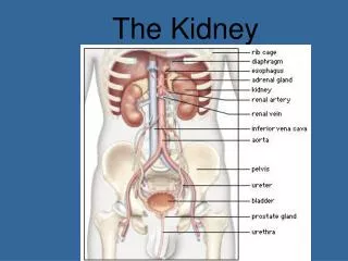
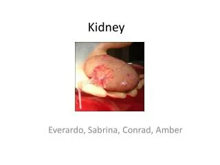
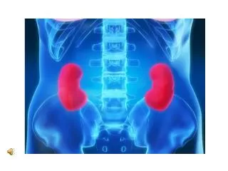
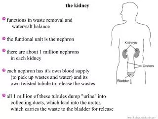
Anatomy of a kidney Renal Renal
Function of kidney • Renal artery takes blood into kidney • Blood drains away from each kidney by the renal vein • Urine is the fluid produced by kidneys and consists of water and dissolved waste products which have been removed from the bloodstream • Urine collects within each renal pelvis • Renal pelvis drains urine into each ureter, which then takes the urine to the urinary bladder
Glomerulus and associated nephron • Each kidney has 1.25 million filtering units known as nephrons • Each nephron consists of: • Capillary bed, called a glomerulus, which filters various substances from the blood • Capsule surrounding the glomerulus called Bowman’s capsule • A small tube (tubule) that extends from Bowman’s capsule and has parts named • Proximal convoluted tubule • Loop of Henle • Distal convoluted tubule • Second capillary bed called the peritubular capillary bed which surrounds the three-part tubule mentioned above
Ultrafiltration • Term used to describe the process by which various substances are filtered through the glomerulus under the unusually high blood pressure in this capillary bed • Walls of the capillaries have very small slits called fenestrations which open when blood pressure is increased (due to smaller diameter of efferent arteriole) • Fluid which is ultrafiltered then passes through the basement membrane which helps prevent large molecules like proteins from becoming a part of the filtrate • The filtrate next enters the proximal convoluted turuble • Blood cells, proteins, and other molecules which did not become a part of the filtrate exit Bowman’s capsule by way of the efferent tubule
Reabsorption • Filtrate that leaves Bowman’s capsule contains many substances that the body cannot afford to lose in the urine, such as water, salt ions, and glucose • Substances need to be reabsorbed into the bloodstream • Most reabsorption takes place from the proximal convoluted tubule • Substances leave the tubule filtrate and are taken back into the bloodstream by way of the peritubular capillary bed
Proximal convoluted tubule • Wall is a single cell thick • Composed of a ring of cells • Interior of tube is called the lumen • Filtrate flows through the lumen • Inner portion of the tubule cells has microvilli which increase the surface area for reabsorption
Transport mechanisms of reabsorption • Salt ions: (Na+, Cl-, K+) activelytransported into the tubule cells and then into the intercellular fluid outside the tubule; then taken into the peritubular capillary bed • Water: water follows salt ions by osmosis • Glucose: in a properly functioning nephron, all the glucose that is in the glomerular filtrate is reabsorbed in the bloodstream by active transport; otherwise, only 50% is reabsorbed
Osmoregulation • Body’s response mechanisms which attempt to maintain homeostatic levels of water • Total volume of water eliminated depends on: • Total volume of water ingested recently (as liquid and in foods) • Perspiration rate (influenced by exercise and environmental temperature) • Ventilation rate (loss of water through exhalation)
Loop of Henle • Much of the water in the original filtrate remains even after the filtrate has left the proximal convoluted tubule • Water and dissolved solutes enter the descending loop of Henle which is permeable to water, but relatively impermeable to salt ions • The filtrate enters the ascending portion of the loop of Henle where the tubule is relatively impermeable to water, but permeable to salt ions • Loop of Henle extends down into the medulla region and creates a hypertonic environment in the medulla • The filtrate that moves up the ascending loop and into the distal convoluted tubule is relatively hypotonic
ADH and the collecting duct • Collecting duct is differentially permeable to water • Its permeability depends on the presence or absence of antidiuretic hormone (ADH) • ADH is secreted by posterior pituitary gland, circulates in bloodstream, and targets the kidney collecting ducts • If ADH is present, collecting duct becomes more permeable water and water moves out of duct and into the medulla intercellular fluid then into peritubular capillary bed • If ADH is not present, collecting duct becomes impermeable to water and water stays in the collecting duct and the urine is more dilute
Logic behind each change • Proteins are too large to fit through the basement membrane within the glomerulus. Thus proteins do not become a part of the filtrate or urine. • Glucose does become a part of the filtrate, but active transport takes 100% of the glucose back into the peritubular capillary bed. In a healthy person, no glucose appears in the urine. • Urea is not toxic unless its concentration is too high in the blood plasma. The very high concentration of urea in urine is due to the reabsorption of water.
Glucose in urine of untreated diabetics • Active transport mechanisms have a maximum rate at which they can move substances • When the maximum threshold concentration of glucose is exceeded, the active transport cannot move all the glucose back into the bloodstream • Some glucose, therefore, remains in the urine