Download
1 / 17
190 likes | 350 Vues
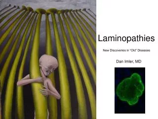
Laminopathies. New Discoveries in “Old” Diseases. Dan Imler, MD. Case Presentation. 13 mo M comes to her PMD’s office Parents Concerns Always has been small (ht and wt) and a poor feeder Has not developed any teeth yet Odd looking nails Always had a strange cry, Dad calls it “Girlie”

E N D
Laminopathies New Discoveries in “Old” Diseases Dan Imler, MD
Case Presentation • 13 mo M comes to her PMD’s office • Parents Concerns • Always has been small (ht and wt) and a poor feeder • Has not developed any teeth yet • Odd looking nails • Always had a strange cry, Dad calls it “Girlie” • Hasn’t seen a physician since 4 mo • Ex FT, no complications • Normal neonatal period • No hospitalizations • Developmentally Normal • No significant Family Hx other than adult CV dz and hypercholesterolemia
Case Presentation • Physical Exam (pertinent findings, all others normal) • Wt 8kg (<3%) | Ht 70cm (<3%) • 3-4 lesions on trunk and extremities of indurated, shiny inelastic skin. Skin generally loose with loss of subcutaneous fat particularly over hands and feet. Freckle-like hyperpigmentation on face and forearms. • Thin terminal hairs. Dystrophic nails • Pointy nose, thin lips, prominent eyes • Mild micrognathia, large anterior fontanel • Thin limbs with prominent joints • Pyriform (pear shaped) thorax • Hip “Clunk” on O & B • Very small nipples
Case Presentation • Test Results • All labs normal except -> low HDL • Brain MRI – diffuse early signs of cerebrovascular occlusive disease • Plain Films / CT – Diffuse osteopenia, Acroosteolysis (distal bone resorption) of the phalanges and distal clavicles, “Fish-Mouth” vertebral bodies, Coxa valga, attenuated cortical bone, wide metaphyses, normal bone age. • Skin Biopsy – epidermis moderately acanthotic. Deposition of thickened, homogenized collagen in the subcutatious tissue. Mild perivascular infiltrate. Increased mucopolysaccharides • Increased levels of hyaluronic acid • LMNA gene positive (Testing done at Brown)
Hutchinson-Gilford Progeria • Progeria – Greek geras – old age • Patients with HPGS develop accelerated atherosclerosis of cerebral and coronary arteries, however the only lipid abnormality associated with the disease is decreased HDL • Patients exhibit clinical signs of accelerated aging including loss of subcutanious fat and muscle, skin atrophy, osteoporosis, arthritis, poor growth and alopecia. • However: patients DO NOT develop other disease processes associated with aging such as increased tumor formation, cataract development or senility -> Segmental Progeroid Syndrome
Hutchinson-Gilford Progeria • 80% of cases the gene defect responsible for HGPS is a single spontaneous mutation in codon 608 of the LMNA gene, which encodes both lamin A and lamin C
Hutchinson-Gilford Progeria • Most mortality due to atherosclerosis • Average life expectancy 13 yrs (7-27) • 97% White population (unknown reason) • 1.5 : 1 Male to Female • Clinically HGPS usually not evident at birth and presenting sx usually seen 6-12 mo usually dx from FTT work-up • Normal intelligence • Emotionally similar to age matched peers, however they have been seen to be keenly aware of their different appearance and remain reserved in the company of strangers. In presence of family-friends, normal social interactions.
Hutchinson-Gilford Progeria • Ageing • Increased Hyaluronic acid only elevated in one other condition – Werner syndrome, a disease with later onset premature ageing • Hyaluronic acid and other gylcosaminoglycans increase in 5th-7th decades of life. 3x higher in progeriaic children • HA is important for morphological development of cells, especially blood vessels which may be the reason for sclerodermatous changes in skin. • Cells from progeria have: reduced mitotic activity, DNA-synthesis, cloning efficiency, ability to repair after irradiation and ? Reduced number of cell divisions.
Treatment • New Clinical treatment trial • Farnesyltransferase inhibitors (FTIs) • The basic principle is the product of the mutated LMNA gene is a malfunctioning protein Prelamin A. Prelamin A has a farnesyl group attached to it which allows it to anchor itself to the nuclear rim. In normal Prelamin A this farnesyl group is removed, however in progeria the persistent molecule causes nuclear abnormalities. Farnesyltransferase inhibitors act to remove this farnesyl group and have been shown in mouse cells to cause normalization of nuclear shape.
Treatment • UCLA has two types of progeria mouse models • Recently they treated them with Farnesyltransferase inhibitors (via drinking water) and showed • Normalization of Nuclear morphology • Reduced Bone Fractures • Increased weight gain • Delayed occurrence of disease • Increased life spans • No data yet on CV disease
Laminopathies • Lamins are intermediate filament proteins that form the nuclear lamina scaffold underneath the nuclear envelope in animal cells. They are attached to the nuclear envelope membrane via farnesyl anchors and interaction with inner nuclear membrane proteins such as lamin B receptor and emerin. The nuclear lamina appears to be an adaptation to mobility in animals as sessile organisms such as plants or fungi do not have lamins and the symptoms of many laminopathies include muscle defects. Mutations in these genes might lead to defects in filament assembly and/or attachment to the nuclear envelope and thus jeopardize nuclear envelope stability in physically stressed tissues such as muscle fibers, bone, skin and connective tissue. • Messenger RNA produced from the LMNA gene undergoes alternative splicing and is translated into lamins A and C. Lamin A undergoes farnesylation to attach a membrane anchor to the protein. This version of the protein is also referred to as prelamin A. Farnesylated prelamin A is further processed into mature lamin A by a metalloproteinase removing the last 15 amino acids and its farnesylated cysteine. This allows lamin A to dissociate from the nuclear envelope membrane and fulfill nuclear functions. Mutations causing laminopathies interfere with these processes on different levels.
Laminopathies • Nonsense and missense mutations • Missense mutations in the lamin A/C rod and tail domains are the cause for a wide array of genetic disorders, suggesting that lamin A/C protein contains distinct functional domains that are essential for the maintenance and integrity of different cell lineages. Interaction between lamin A and the nuclear envelope protein emerin appears to be crucial in muscle cells, with certain mutations in lamin mimicking mutations in emerin and causing Emery-Dreifuss muscular dystrophy. Different mutations lead to dominant-negative and recessive alleles. Mutations in the lamin rod domain leading to mislocalization of both lamin A and emerin occur in patients with autosomal dominant forms of muscular dystrophy and cardiomyopathy. • Most lamin B mutations appear to be lethal with mutations in lamin B1 causing death at birth in mice. In 2006, lamin B2 missense mutations were identified in patients with acquired partial lipodystrophy.
Laminopathies • Splicing defects • Mutations causing progeria are defective in splicing LMNA mRNA, therefore producing abnormal lamin A protein, also known as progerin. The mutations activate a cryptic splice site within exon 11 of the gene, thereby causing the deletion of the processing site on prelamin A. This results in an accumulation of progerin that is unable to mature into lamin A, leading to misshapen nuclei. Missplicing also leads to the complete or partial loss of exon 11 and results in a truncated prelamin A protein in the neonatal lethal tight skin contracture syndrome.