Hydrogen sulfide and meningitis
20 likes | 147 Vues
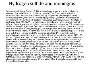
This supplemental digital content presents serial MRI images illustrating changes in the apparent diffusion coefficient (ADC) over time in a patient with meningitis linked to hydrogen sulfide exposure. The ADC variations are color-coded, indicating decreasing (more blue, signifying more cytotoxic edema) or increasing (more red) levels, relative to the admission study. A ventriculoperitoneal shunt in the right hemisphere caused artifacts; only changes in the left hemisphere are displayed. These changes are representative of global alterations, with contiguous spread evident in bilateral cortices and deep gray nuclei, inconsistent with a vascular distribution.

Hydrogen sulfide and meningitis
E N D
Presentation Transcript
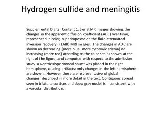
Hydrogen sulfide and meningitis Supplemental Digital Content 1. Serial MR images showing the changes in the apparent diffusion coefficient (ADC) over time, represented in color, superimposed on the fluid attenuated inversion recovery (FLAIR) MRI images. The changes in ADC are shown as decreasing (more blue, more cytotoxic edema) or increasing (more red) according to the color scales shown at the right of the figure, and computed with respect to the admission study. A ventriculoperitoneal shunt was placed in the right hemisphere, causing artifacts; only changes in the left hemisphere are shown. However these are representative of globalchanges, described in more detail in the text. Contiguous spread seen in bilateral cortices and deep gray nuclei is inconsistent with a vascular distribution.