Download
1 / 56
590 likes | 776 Vues
Animal models for muscular dystrophy. Vincenzo Nigro Laboratorio di genetica - Dipartimento di Patologia Generale, Seconda Università degli Studi di Napoli Telethon Institute of Genetics and Medicine, Napoli. muscular dystrophy.

E N D
Animal models for muscular dystrophy Vincenzo Nigro Laboratorio di genetica - Dipartimento di Patologia Generale, Seconda Università degli Studi di Napoli Telethon Institute of Genetics and Medicine, Napoli
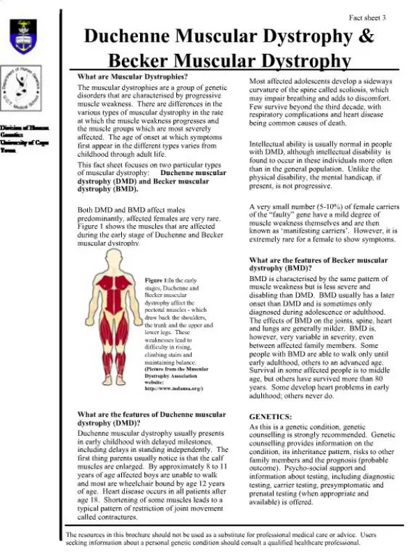
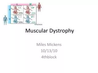
muscular dystrophy • MD is a general term that describes a group of inherited and gradually debilitating myogenic disorders • progressive muscle weakness affecting patients since from young age and can lead to early death • pattern of inheritance can be X-linked recessive (DMD/BMD), autosomal dominant (LGMD1), or autosomal recessive (LGMD2) • Some underlying genetic defects are well known, others are orphan diseases
dystrophin mutations • DMD Duchenne Muscular Dystrophy - 1/3,500 boysOnset -- Early childhood - about 2 to 6 yearsSymptoms -- Generalized weakness and muscle wasting affecting limb and trunk muscles first. Calves often enlargedProgression -- Disease progresses slowly but will affect all voluntary muscles. Survival possible beyond late twenties • BMD Becker Muscular Dystrophy - 1/10,000 boysOnset -- Adolescence or adulthoodSymptoms -- Almost identical to Duchenne but often much less severe. Can be significant heart involvementProgression -- Slower and more variable than Duchenne with survival well into mid to late adulthood
weakness Proximal weakness: the most common site of weakness in a myopathic disorder • Lower extremities • difficulty climbing stairs • arising from a low chair or toilet • getting up from a squatted position • Upper extremities • trouble lifting objects over the head • brushing the hair
fatigue • Much less useful “negative” symptom (non-specific) • Many patients who complain of diffuse global "weakness" or fatigue do not have a disorder of muscle • Abnormal fatigability after exercise: • metabolic and mitochondrial myopathies • define the duration and intensity
disease progression • muscle tissue represent about 40% of the total body mass • respiratory failure can be the cause of premature death as well as heart failure • patients suffer from asymmetries in strength between reciprocal muscles that cause widespread joint and spine deformities requiring timely orthopaedic surgery
Distribution of muscle involvement • CK (50x to 1.000x) • LDH5, ALT, AST, aldolase increase • Clinical diagnosis of LGMD is often made when disease has no apparent X-linked inheritance • LGMD are classified as severe (Duchenne-like) or mild (Becker-like), depending on the rate of progression and the age of wheelchair confinement
management > treatment > therapy > cure Animal models disease should be comparable to human defects Profoundly studied in all pathological characteristics Should allow a reliable prediction of the response
The genetic basis of the disease should be the same as human disease • Reiterate key hallmarks of the human disease • Animals commercially available, easy to maintain • Animal disease well characterized, with abundant literature • Robust phenotype that is reproducible over generations
mdx (X-chr MD) mouseC57BL/10ScSn-Dmdmdx • mdx is the best characterized mouse model for muscular dystrophy (>1,700 papers) since 1984 • mdx has a spontaneous nonsense mutation (stop) in exon 23 of the dystrophin gene and does not produce dystrophin • absence of dystrophin reduces the DGC at the sarcolemma
mdx mouse may be DMD mouse? • mdx shows signs of MD during first 6 weeks of life which results in an increase of the newly differentiated myofibers • it has muscle regeneration with an expansion of the satellite cell population and muscle hypertrophy • Centralized nuclei (50-60%), heterogeneity in fiber size • Necrosis at early stages, but decreases after 60 days • Plasma creatine kinase is 5.000-12.000 U/L • the most affected muscle (diaphragm) reproduces the degenerative changes of MD
4 weeks of age, soleus muscle mdx C57
mdx mouse is a bad DMD mouse model • Fibrosis is only in diaphragm • Absolute muscle force of limb muscles remains similar to unaffected mice • Lifespan is shorter but no so much (-19% in males) • it has muscle regeneration with an expansion of the satellite cell population and muscle hypertrophy • mdx lacking the muscle-specific transcription factor MyoD or myocyte nuclear factor (expressed in the satellite cells) show more severe MD
Double mouse mutants utrophin/dystrophin • Utrophin is a developmentally regulated protein, an autosomal homologue to dystrophin • utrophin is overexpressed when dystrophin is absent • the utrn−/−/mdx mice are severely affected • reduced lifespan • severe muscle weakness with joint contractures, growth retardation, and cardiomyopathy • the phenotype is ameliorated by skeletal-muscle specific expression of utrophin
Dp260 = retinal Dp140 = central nervous system and kidney Dp116 = Schwann cells Dp71 = high levels, but not in muscle
mdx52 mouse • mdx52 is dystrophin KO mouse • mdx has a deletion in exon 52 of the dystrophin gene and does not produce dystrophin • in contrast to mdx, this mouse cannot produce also Dp260(ret) and Dp140(CNS), maintaining Dp116 (S) and Dp71 • it is very similar to mdx mouse with the absence of dystrophin that reduces the DGC at the sarcolemma, but has no cardiomyopathy
mdx2cv-5cv mice • they were generated by chemical mutagenesis using N-ethyl-nitrosurea • mdx 2cv lacks dys, Dp260 • mdx 3cv lacks dys, Dp260, Dp140, Dp116 and Dp71 • mdx 4cv lacks dys, Dp260 and Dp140 • mdx 5cv lacks only dystrophin • phenotypes are very similar to mdx mouse and no phenotype worsening
targeted inactivation of Dp71 only • there is a mouse that cannot produce Dp71 only • it has normal phenotype
Dystrophin revertant fibres and transcripts in mdx mouse muscle Fall et al. Genetic Vaccines and Therapy 2006 4:3
the "humanized" hDMD mouse • “humanised” DMD (hDMD) mice carry an integrated and functional copy of the full-length human DMD gene • it serves to test the “exon skipping strategy” that is a sequence-specific therapeutic approach
human sequence-specific DMD exon skipping in vivo • the hDMD mouse model allows the direct testing of human-specific AONs and target sequences in a mouse experimental background • the induction of specific skipping of the hDMD exons 44, 46, and 49, whilst the endogenous mouse transcripts are not affected [Bremmer-Bout et al., Mol. Ther. 2004] • this underlines that AONs, based upon specific design, can be highly sequence-specific small molecule drugs.
RT-PCR analyses using either mouse- or human-specific primers show correct transcription of the human DMD gene in muscle tissue hDMD mouse mRNA
expression of human dystrophin in skeletal muscle detected by IF using the human-specific Ab MANDYS106Dys2 reacts with both human and mouse dystrophin hDMD mouse IF
Online Mendelian Inheritance in Animals (OMIA)is a database of genes, inherited disorders and traits in more than 135 animal species (other than human and mouse, which have their own resources). The database contains textual information and references, as well as links to relevant PubMed and Gene records at the NCBI
Simple search for: "muscular dystrophy" 10 records found OMIA 000679 Muscular dystrophy in Gallus gallus (chicken)Sub-type: Abnormal muscle; AM Genes: WWP1 OMIA 000679 Muscular dystrophy in Canis familiaris (dog) OMIA 000679 Muscular dystrophy in Ovis aries (sheep) OMIA 000679 Muscular dystrophy in Meleagris gallopavo (turkey) OMIA 000679 Muscular dystrophy in Mustela lutreola (European mink) OMIA 000679 Muscular dystrophy in Felis catus (cat) OMIA 001081 Muscular dystrophy, Duchenne and Becker types in Felis catus (cat)Genes: DMD OMIA 001081 Muscular dystrophy, Duchenne and Becker types in Canis familiaris (dog)Sub-type: X-linked muscular dystrophy OMIA 000681 Muscular dystrophy, dysphagia-associated in Canis familiaris (dog) OMIA 000828 Progressive muscular dystrophy in Mustela lutreola (European mink European mink)
Golden retriever dog with muscular dystrophy (GRMD) GRMD arises from a mutation in the acceptor splice site of intron 6 of the dystrophin gene Skipping of exon 7 disrupts the mRNA reading frame and results in premature termination of translation
Golden retriever dog with muscular dystrophy (GRMD) complete absence of the dystrophin, early and severe muscle degeneration with reduction of motility and walking ability Death usually occurs at about 1 year of age as a result of failure of respiratory muscles
dystrophic Golden Retriever dog • gradual weakness and loss of muscle mass • development of contractures, skeletal deformities • significant phenotypic variability among litters
Spitz dogs • Becker-like dystrophy with a truncated form of dystrophin was recently identified in a family of Japanese Spitz dogs
LGMD forms • LGMD have a highly variable onset and progression, but the unifying theme is the proximal muscle involvement • The a. dominant forms (LGMD1) are generally milder and relatively rare representing less than 10% of all LGMD • The a. recessive forms (LGMD2) are much more common, having a cumulative prevalence of 1:14,000-1:20,000 with some differences among countries, depending on the carrier distribution and the degree of consanguinity • There are, however, at least 25% of families who can be excluded from any known locus and 40% of typical LGMD cases with no mutation in any known gene
Autosomal dominant LGMD1A 5q31.2 myotilin (Hauser, 2000) LGMD1B 1q21 lamin A/C (Bonne, 1999) LGMD1C 3p25.3 caveolin 3 (Minetti, 1997) LGMD1D 6q22 ? LGMD1E 7q35 ? LGMD1F 7q31.1 ? LGMD1G 4p21 ? Autosomal recessive LGMD2A 15q15 calpain 3 (Richard, 1995) LGMD2B 2p13.2 dysferlin (Bashir, Liu, 1998) LGMD2C 13q12 g-sarcoglycan (Noguchi, 1995) LGMD2D 17q21.33 a-sarcoglycan (Roberds, 1994) LGMD2E 4q12 b-sarcoglycan (Bonnemann, Lim, 1995) LGMD2F 5q33 d-sarcoglycan (Nigro, 1996) LGMD2G 17q12 telethonin (Moreira, 2000) LGMD2H 9q33.1 TRIM 32 (Frosk, 2002) LGMD2I 19q13.3 FKRP (Brockington, 2001) LGMD2J 2q24.3 titin (Udd, 2002) LGMD2K 9q34.1 POMT1 (Balci, 2005) LGMD2L 9q31 fukutin (Godfrey, 2006) LGMD2M 1p34.1 POMGnT1 (Clement, 2008) LGMD2N 14q24 POMT2 (Biancheri, 2007) LGMD2O 11p13-p12 ? (Jarry, 2007)
Dy/dy dy2J/dy2J • Two mouse models for laminin-α2 deficiency were identified in the Jackson Laboratories (http://www.jax.org/) • dy/dy (dystrophia-muscularis) mouse • allelic dy2J/dy2J mouse • Both mice are models for merosin-deficient Congenital MD (CMD1A) • Neither of these mouse models exhibits a complete deficiency of laminin α2 chain
alfa-syntrophin -/- mouse • no defect in muscle • nNOS and aquaporin-4 are displaced, like in mdx • aquaporin KO are also normal • nNOS KO are normal • nNOS(-/-)/mdx are = mdx
alfa-dystrobrevin -/- mouse • maintain the expression of DGC at the sarcolemma • mild muscular dystrophy but not yet in humans • is affected DGC complex signaling? • nNOS and aquaporin-4 are displaced, like in mdx • aquaporin KO are also normal
Mutation in any of the sarcoglycan genes produces a secondary loss of the other components
LGMD2C, 2D, 2E, 2F with sarcoglycan gene mutations • Mutation in any of the sarcoglycan genes produces a phenotype very similar to DMD/BMD • Onset between 7-19 years, variable progression with some patients that never loss deambulation and other that are more severely affected, also with identical mutations • Atrofic muscular dystrophy. Calf hypertrophy. It may involve distal muscles. Little shoulder girdle involvement. Heart is seldom involved. CK is very high
N C C N sarcoglycan family members protein aa MW expression ex. chrom a-sarcoglycan 387 50 muscle 10 17q12 e-sarcoglycan 413 52 ubiquit. 12 7q21 g-sarcoglycan 291 35 muscle 8 13q12 d-sarcoglycan 290 35 muscle 9 5q33 z-sarcoglycan 299 36 brain 9 8p22 b-sarcoglycan 318 43 muscle 6 4q12
alternative sarcoglycan complexes a b g d e b z d e b g d
Spe I Hind III Hind III 1.3 kb 9.5 kb NEO b gal Spe I Hind III Eco RV Eco RV Eco RV Hind III intron 9 e-SG intron 5 e-SG exx. 7- 9 ex. 6 Spe I Hind III Hind III intron 9 e-SG intron 5 e-SG NEO b gal We deleted exons 6-9 of e-sarcoglycan encoding Cys-rich and transmembrane domains K.O. of the e-sarcoglycan gene vector target locus K.O.