1 / 26
260 likes | 430 Vues
Our Vein treatment procedures have the least recovery time & give long term outstanding results. All our Vein Centers have in-house diagnostic Ultrasound and Colour Doppler facilities that gives us a precise roadmap to plan your Varicose veins treatment.

E N D
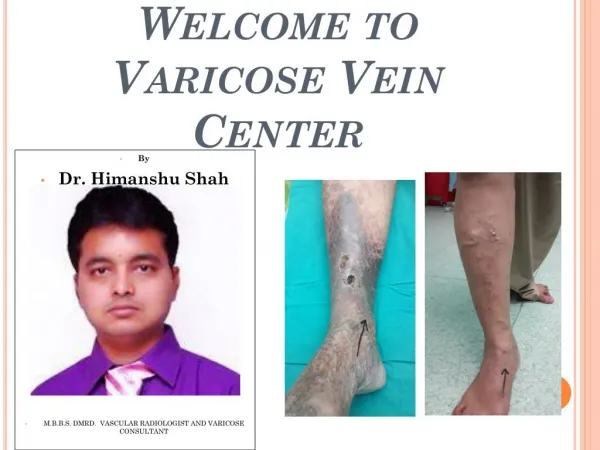
Welcome toVaricose Vein Center • By • Dr. Himanshu Shah • M.B.B.S. DMRD. VASCULAR RADIOLOGIST AND VARICOSE CONSULTANT
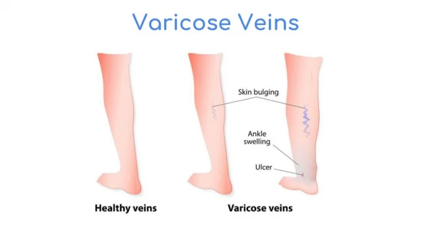
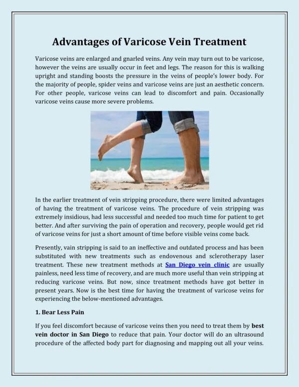
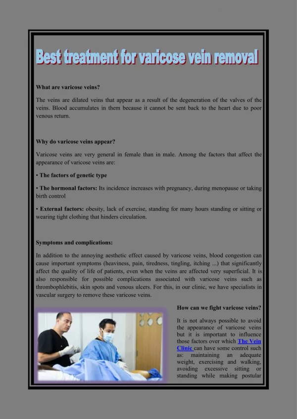
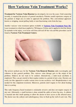
DR. HIMANSHU SHAH M.B.B.S.D.M.R.D., VARICOSEVEIN CONSULTANT Varicose veins • Varicose veins are veins that have become enlarged and twisted. The term commonly refers to the veins on the leg,although varicose veins can occur elsewhere. Veins have pairs of leaflet valves to prevent blood from flowing backwards (retrograde flow or venous reflux). Leg muscles pump the veins to return blood to the heart (the skeletal-muscle pump), against the effects of gravity. When veins become varicose, the leaflets of the valves no longer meet properly, and the valves do not work (valvular incompetence). This allows blood to flow backwards and they enlarge even more. Varicose veins are most common in the superficial veins of the legs, which are subject to high pressure when standing. Besides being a cosmetic problem, varicose veins can be painful, especially when standing. Severe long-standing varicose veins can lead to leg swelling, venous eczema, skin thickening (lipodermatosclerosis) and ulceration. Life-threatening complications are uncommon, but varicose veins may be confused with deep vein thrombosis, which may be life-threatening
ulcer varicose veins in legs • Non-surgical treatments include sclerotherapy, elastic stockings, leg elevation and exercise. The traditional surgical treatment has been vein stripping to remove the affected veins. Newer, less invasive treatments which seal the main leaking vein are available. Alternative techniques, such as ultrasound-guided foam sclerotherapy, radiofrequency ablation and endovenous laser treatment, are available as well. Because most of the blood in the legs is returned by the deep veins, the superficial veins, which return only about 10% of the total blood of the legs, can usually be removed or ablated without serious harm. • Secondary varicose veins are those developing as collateral pathways, typically after stenosis or occlusion of the deep veins, a common sequel of extensive deep venous thrombosis (DVT). Treatment options are usually support stockings, occasionally sclerotherapy and rarely, limited surgery.
DR. HIMANSHU SHAH M.B.B.S.D.M.R.D., VARICOSEVEIN CONSULTANT Signs and symptoms • Aching, heavy legs (often worse at night and after exercise). • Appearance of spider veins (telangiectasia) in the affected leg. • Ankle swelling, especially in evening. • A brownish-yellow shiny skin discoloration near the affected veins. • Redness, dryness, and itchiness of areas of skin, termed stasis dermatitis or venous eczema, because of waste products building up in the leg.
DR. HIMANSHU SHAH M.B.B.S.D.M.R.D., VARICOSEVEIN CONSULTANT • Cramps may develop especially when making a sudden move as standing up. • Minor injuries to the area may bleed more than normal or take a long time to heal. • In some people the skin above the ankle may shrink (lipodermatosclerosis) because the fat underneath the skin becomes hard. • Restless legs syndrome appears to be a common overlapping clinical syndrome in patients with varicose veins and other chronic venous insufficiency. • Whitened, irregular scar-like patches can appear at the ankles. This is known as atrophie blanche.
DR. HIMANSHU SHAH M.B.B.S.D.M.R.D., VARICOSEVEIN CONSULTANT Complications Most varicose veins are reasonably benign, but severe varicosities can lead to major complications, due to the poor circulation through the affected limb.
Pain, tenderness, heaviness, inability to walk or stand for long hours, thus hindering work • Skin conditions / Dermatitis which could predispose skin loss • Skin ulcers especially near the ankle, usually referred to as venous ulcers. • Development of carcinoma or sarcoma in longstanding venous ulcers. Over 100 reported cases of malignant transformation have been reported at a rate reported as 0.4% to 1%. • Severe bleeding from minor trauma, of particular concern in the elderly. • Blood clotting within affected veins, termed superficial thrombophlebitis. These are frequently isolated to the superficial veins, but can extend into deep veins, becoming a more serious problem. • Acute fat necrosis can occur, especially at the ankle of overweight patients with varicose veins. Females are more frequently affected than males.
DR. HIMANSHU SHAH M.B.B.S.D.M.R.D., VARICOSEVEIN CONSULTANT Diagnosis Clinical tests Investigations • Clinical tests that may be used include: • Trendelenburg test–to determine the site of venous reflux and the nature of the sapheno femoral junction • Multiple tournique test–to more accurately localize the site of the venous reflux • Fegan's test–to assess the nature of any perforating vein blow outs • Perthes test–to check the patency of the deep veins • Other more historical/ academic tests include Scwhartz test, and Morrisey's cough impulse test. • Lower limbs venous ultrasonography has replaced most of the rest. • Traditionally, varicose veins were investigated using imaging techniques only if there was a clinical suspicion of deep venous insufficiency, if they were recurrent, or if they involved the sapheno-popliteal junction. This practice is not now widely accepted. Patients with varicose veins should now be investigated using lower limbs venous ultrasonography. The results from a randomised controlled trial on patients with and without routine ultrasound has shown a significant difference in recurrence rate and reoperation rate at 2 and 7 years of follow up.
According to the CEAP classification: C0 –no visible or palpable signs of venous disease C1 –telangectasia or reticular veins C2 –varicose veins. C3 –edema C4a –pigmentation or eczema C4b –lipodermatosclerosis, atrophie blanche C5 –healed venous ulcer C6 –active venous ulcer •Each clinical class is further characterised by a subscript depending upon whether the patient is symptomatic (S) or asymptomatic (A) e.g. C2S.
DR. HIMANSHU SHAH M.B.B.S.D.M.R.D., VARICOSEVEIN CONSULTANT Causes Varicose veins are more common in women than in men, and are linked with heredity. Other related factors are pregnancy, obesity, menopause, aging, prolonged standing, leg injury and abdominal straining. Varicose veins are unlikely to be caused by crossing the legs or ankles. Less commonly, but not exceptionally, varicose veins can be due to other causes, as post phlebitic obstruction or incontinence, venous and arteriovenous malformations. More recent research has shown the importance of pelvic vein reflux (PVR) in the development of varicose veins. Hobbs showed varicose veins in the legs could be due to ovarian vein reflux and Lumley and his team showed recurrent varicose veins could be due to ovarian vein reflux. Whiteley and his team reported that both ovarian and internal iliac vein reflux causes leg varicose veins and that this condition affects 14% of women with varicose veins or 20% of women who have had vaginal delivery and have leg varicose veins. In addition evidence suggests that failing to look for, and treat pelvic vein reflux can be a cause of recurrent varicose veins.
There is increasing evidence for the role of incompetent Perforator veins (or "perforators") in the formation of varicose veins and recurrent varicose veins. Varicose veins could also be caused by hyperhomocysteinemia in the body, which can degrade and inhibit the formation of the three main structural components of the artery: collagen, elastin and the proteoglycans. Homocysteine permanently degrades cysteinedisulfide bridges and lysineamino acid residues in proteins, gradually affecting function and structure. Simply put, homocysteine is a 'corrosive' of long-living proteins, i.e. collagen or elastin, or lifelong proteins, i.e. fibrillin. These long-term effects are difficult to establish in clinical trials focusing on groups with existing artery decline. Klippel-Trenaunay syndrome and Parkes-Weber syndrome are relevant for differential diagnosis. Another cause is chronic alcohol consumption due to the vasodilatating side effect in relation to gravity and blood viscosity.
DR. HIMANSHU SHAH M.B.B.S.D.M.R.D., VARICOSEVEIN CONSULTANT Treatment Treatment can be either conservative or active. Active treatments can be divided into surgical and non-surgical treatments. Newer methods including endovenous laser treatment, radiofrequency ablation and foam sclerotherapy appear to work as well as surgery for varices of the greater saphenous vein.
DR. HIMANSHU SHAH M.B.B.S.D.M.R.D., VARICOSEVEIN CONSULTANT Conservative The National Institute for Health and Clinical Excellence (NICE) produced clinical guidelines in July 2013 recommending that all people with symptomatic varicose veins (C2S) and worse should be referred to a vascular service for treatment. Conservative treatments such as support stockings should not be used unless treatment was not possible.
The symptoms of varicose veins can be controlled to an extent with the following: Elevating the legs often provides temporary symptomatic relief. Advice about regular exercise sounds sensible but is not supported by any evidence. The wearing of graduated compression stockings with variable pressure gradients (Class II or III) has been shown to correct the swelling, nutritional exchange, and improve the microcirculation in legs affected by varicose veins. They also often provide relief from the discomfort associated with this disease. Caution should be exercised in their use in patients with concurrent arterial disease.
DR. HIMANSHU SHAH M.B.B.S.D.M.R.D., VARICOSEVEIN CONSULTANT The wearing of intermittent pneumatic compression devices have been shown to reduce swelling and increase circulation Diosmin/Hesperidine and other flavonoids. Anti-inflammatory medication such as ibuprofen or aspirin can be used as part of treatment for superficial thrombophlebitis along with graduated compression hosiery – but there is a risk of intestinal bleeding. In extensive superficial thrombophlebitis, consideration should be given to anti-coagulation, thrombectomy or sclerotherapy of the involved vein.Topical gel application, helps in managing symptoms related to varicose veins such as inflammation, pain, swelling, itching and dryness. Topical application-noninvasive has patient compliance.
DR. HIMANSHU SHAH M.B.B.S.D.M.R.D., VARICOSEVEIN CONSULTANT Surgical Surgeries have been performed for over a century, from the more invasive saphenous stripping, to less invasive procedures like ambulatory phlebectomy and CHIVA. Stripping Other • Stripping consists of removal of all or part the saphenous vein (great/long or lesser/short) main trunk. The complications include deep vein thrombosis (5.3%), pulmonary embolism (0.06%), and wound complications including infection (2.2%). There is evidence for the great saphenous vein regrowing after stripping. For traditional surgery, reported recurrence rates, which have been tracked for 10 years, range from 5–60%. In addition, since stripping removes the saphenous main trunks, they are no longer available for use as venous bypass grafts in the future (coronary or leg artery vital disease) • Other surgical treatments are: • Ambulatory phlebectomy • Veinligation is done at sephenofemoral junction after ligating the tributeries at sephanofemoral junction without stripping the long sephenous vein provided the perforater veins are competent and absent DVT in the deep veins.With this method long sephenous vein is preserved. • Cryosurgery- A cryoprobe is passed down the long saphenous vein following saphenofemoral ligation. Then the probe is cooled with NO2 or CO2 to −85o F. The vein freezes to the probe and can be retrogradely stripped after 5 seconds of freezing. It is a variant of Stripping. The only point of this technique is to avoid a distal incision to remove the stripper.
A 1996 study reported a 76% success rate at 24 months in treating saphenofemoral junction and great saphenous vein incompetence with STS 3% solution. A Cochrane Collaboration review concluded sclerotherapy was better than surgery in the short term (1 year) for its treatment success, complication rate and cost, but surgery was better after 5 years, although the research is weak. A Health Technology Assessment found that sclerotherapy provided less benefit than surgery, but is likely to provide a small benefit in varicose veins without reflux. This Health Technology Assessment monograph included reviews of epidemiology, assessment, and treatment, as well as a study on clinical and cost effectiveness of surgery and sclerotherapy. Complications of sclerotherapy are rare but can include blood clots and ulceration. Anaphylactic reactions are "extraordinarily rare but can be life-threatening," and doctors should have resuscitation equipment ready. There has been one reported case of stroke after ultrasound guided sclerotherapy when an unusually large dose of sclerosant foam was injected.
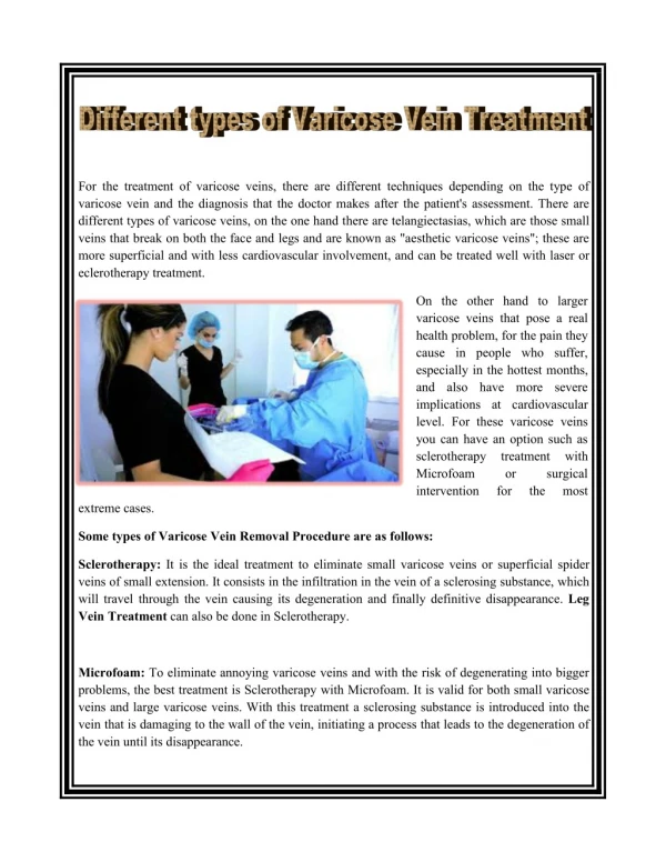
DR. HIMANSHU SHAH M.B.B.S.D.M.R.D., VARICOSEVEIN CONSULTANT Sclerotherapy A commonly performed non-surgical treatment for varicose and "spider" leg veins is sclerotherapy, in which medicine (sclerosant) is injected into the veins to make them shrink. The medicines that are commonly used as sclerosants are polidocanol (POL branded Asclera in the United States, Aethoxysklerol in Australia), sodium tetradecyl sulphate (STS), Sclerodex (Canada), Hypertonic Saline, Glycerin and Chromated Glycerin. STS (branded Fibrovein in Australia) liquids can be mixed at varying concentrations of sclerosant and varying sclerosant/gas proportions, with air or CO2 or O2 to create foams. Foams may allow more veins to be treated per session with comparable efficacy. Their use in contrast to liquid sclerosant is still somewhat controversial. Sclerotherapy has been used in the treatment of varicose veins for over 150 years. Sclerotherapy is often used for telangiectasias (spider veins) and varicose veins that persist or recur after vein stripping.Sclerotherapy can also be performed using foamed sclerosants under ultrasound guidance to treat larger varicose veins, including the great saphenous and small saphenous veins.
DR. HIMANSHU SHAH M.B.B.S.D.M.R.D., VARICOSEVEIN CONSULTANT Endovenous thermal ablation There are three kinds of endovenous thermal ablation treatment possible laser, radiofrequency and steam. The Australian Medical Services Advisory Committee (MSAC) in 2008 determined that endovenous laser treatment/ablation (ELA) for varicose veins "appears to be more effective in the short term, and at least as effective overall, as the comparative procedure of junction ligation and vein stripping for the treatment of varicose veins." It also found in its assessment of available literature, that "occurrence rates of more severe complications such as DVT, nerve injury and paraesthesia, post-operative infections and haematomas, appears to be greater after ligation and stripping than after EVLT". Complications for ELA include minor skin burns (0.4%) and temporary paraesthesia (2.1%). The longest study of endovenous laser ablation is 39 months.
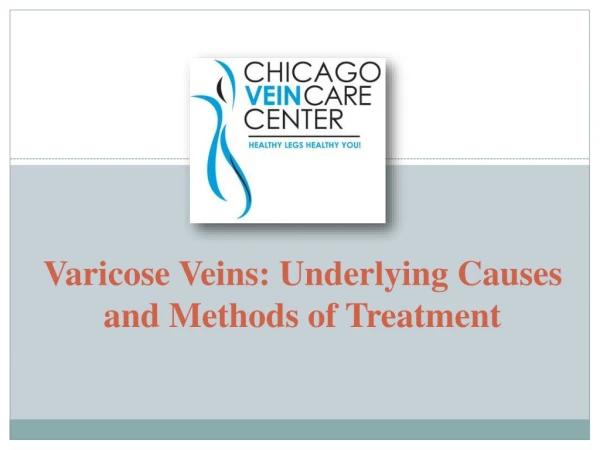
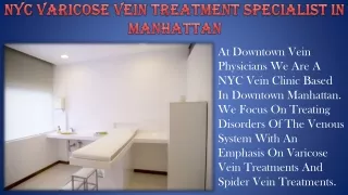
DR. HIMANSHU SHAH M.B.B.S.D.M.R.D., VARICOSEVEIN CONSULTANT BEFORE AFTER BEFORE AFTER
Two prospective randomized trials found speedier recovery and fewer complications after radiofrequency ablation (ERA) compared to open surgery. Myers wrote that open surgery for small saphenous vein reflux is obsolete. Myers said these veins should be treated with endovenous techniques, citing high recurrence rates after surgical management, and risk of nerve damage up to 15%. By comparison ERA has been shown to control 80% of cases of small saphenous vein reflux at 4 years, said Myers. Complications for ERA include burns, paraesthesia, clinical phlebitis and slightly higher rates of deep vein thrombosis (0.57%) and pulmonary embolism (0.17%). One 3-year study compared ERA, with a recurrence rate of 33%, to open surgery, which had a recurrence rate of 23%. Steam treatment consists in injection of pulses of steam into the sick vein. This treatment which works with a natural agent (water) has similar results than laser or radiofrequency. The steam presents a lot of post-operative advantages for the patient (good aesthetic results, less pain, etc.)
DR. HIMANSHU SHAH M.B.B.S.D.M.R.D., VARICOSEVEIN CONSULTANT ELA and ERA require specialized training for doctors and special equipment. ELA is performed as an outpatient procedure and does not require an operating theatre, nor does the patient need a general anaesthetic. Doctors use high frequency ultrasound during the procedure to visualize the anatomical relationships between the saphenous structures. Some practitioners also perform phlebectomy or ultrasound guided sclerotherapy at the time of endovenous treatment. Follow-up treatment to smaller branch varicose veins is often needed in the weeks or months after the initial procedure. Steam is a very promising treatment for both doctors (easy introduction of catheters, efficient on recurrences, ambulatory procedure, easy and economic procedure) and patients (less post-operative pain, natural agent, fast recovery to daily activities). THANK YOU