Download

1 / 34
340 likes | 365 Vues
Explore the confined DNA and RNA packaging in viruses, from scratch virus production, and the biology and history of viruses. Learn about viral genomes, sizes, and life cycles. Dive into the physical principles behind virus creation and understand the complexities of virus infectivity.

E N D
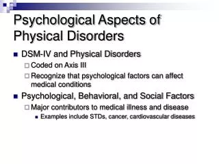
BOULDER 2006: WILLIAM M. GELBART PHYSICAL ASPECTS OF VIRAL INFECTIVITY LECTURE 1: DNA PACKAGING IN VIRUSES DNA is strongly confined; packaged by force LECTURE 2: RNA PACKAGING IN VIRUSES RNA is weakly confined; packaged spontaneously LECTURE 3: MAKING VIRUSES “FROM SCRATCH” Possible for both DNA and RNA cases (The role of membranes)
QUESTIONS WE’LL BE ASKING: WHAT ARE VIRUSES? IN WHAT SENSE ARE VIRUSES ALIVE? HOW IS IT POSSIBLE FOR THEM TO HAVE SO FEW GENES, AND HENCE SUCH A SMALL “PARTS LIST”? WHAT ARE THEIR BASIC/GENERIC “LIFE CYCLES”? AND HOW DO WE UNDERSTAND THEM IN TERMS OF BASIC PHYSICAL PRINCIPLES? WHY ARE MOST VIRUSES SPHERICAL/ICOSAHEDRAL? WHY -- HOW! -- IS IT POSSIBLE TO MAKE INFECTIOUS VIRUSES “FROM SCRATCH”, I.E., FROM PURIFIED COMPONENTS?
HOW BIG IS A VIRUS, AND WHY? *MUST BE SMALL COMPARED TO CELL SIZE (i.e., << a micron) *BUT MUST BE BIG ENOUGH TO ENCLOSE ITS GENOME SO, HOW BIG IS A VIRAL GENOME? How big is a gene ? 1000-base-pair length of DNA has a volume of 1000 x 0.34 nm x p (1.5 nm)2 = 2400 nm3 What about 10 genes? 24000 nm3, suggesting a volume with a radius of 20 nm [VOLUMES SCALE AS CUBES OF LINEAR DIMENSIONS, IMPLYING FACTOR OF 10 RANGE IN GENOME VOLUME GIVES ONLY FACTOR OF TWO CHANGE IN VIRUS SIZE]
ALL VIRUSES ARE ABOUT 50 nm IN DIAMETER….! This is why viruses (unlike bacteria and other infectious microbes): *were notvisible (i.e., were not detectable in the best microscopes available at the time -- one century ago), and *were not filterable in efforts to isolate them: INDEED, VIRUSES -- IN PLANT, ANIMAL, AND BACTERIAL CASES -- WERE DISCOVERED BY INFECTING HOSTS WITH THE FLUIDS COLLECTED FROM FILTERING GROUND-UP LEAVES, BLOOD, AND FECES…FROM INFECTED HOSTS…
SOME HISTORY OF VIRUSES: plant case first to be discovered, early-1890’s (TMV) first to be crystallized, 1935 (TMV) first to be reconstituted, 1955 (TMV) animal case discovered, mid-1890’s (FMDV) cell culture and plaque assays 1952 vaccines (polio), 1950’s; gene delivery… bacterial case discovered, 1915 (“intestinal phage”) phage and the origin of molecular biology, 1937 -- first EM pictures, 1941
d’Herelle’s 1915 discovery of bacteriophage, simultaneous with that of Twort “I made an emulsion of some still bloody stool and filtered it…; to a broth culture of the dysentery bacillus isolated days earlier, I added a drop of the filtrate and spread a drop of this mixture on agar.” “…The next morning, on opening the incubator, I experienced one of those rare moments of intense emotion* which reward the research worker for his pains: at the first glance I saw that the broth culture, which the night before had been very turbid, was perfectly clear -- all the bacteria had vanished.” “…What caused my emotion was that in a flash I had understood: what caused my clear spots was in fact an invisible microbe -- a filterable virus, a virus parasitic on bacteria.” * whilst examining feces in a morgue…!
FIRST EM PICTURE (NOBEL-PRIZE-WINNING) OF PHAGE (1941, RUSKA) CURRENT EM PICTURES OF PHAGE (2005, EVILEVITCH)
T5 bacteriophage “infecting” a lecithin vesicle reconstituted with a few receptor (FhuA) molecules, and full of spermine Lambert, Letellier, Gelbart and Rigaud, PNAS 97, 7248 (2000)
ANIMAL CELL “LIFE CYCLE” *Entry involves receptor-mediated binding/endocytosis or fusion *Whole viral particle enters cell *New virions leave via “budding”
plasmodesmata *Cells are each surrounded by a rigid (cellulose) wall, which must be “broken” (e.g., by abrasion) in order for viral particles to enter *Consequently, a large number of viral particles enter the celland without the need for a receptor-mediated process *Replicated virions leave cell through plasmodesmata
LET’S PURSUE THE LIFE CYCLE OF BACTERIAL VIRUSES… Q: WHAT IS (PHYSICALLY) RESPONSIBLE FOR INJECTIONOF THE MANY-MICRON LONG DNA? A: THE PRESSURE -- ENERGY DENSITY -- INSIDE THE VIRAL CAPSID WHAT IS THE ORIGIN OF THIS STRESS? HOW CAN ONE CALCULATE AND MEASURE FORCES AND PRESSURES OF THIS KIND? STRESS IMPLIES THAT (FREE) ENERGY HAS BEEN STORED: WHAT PERFORMS THE WORK OF ACCOMPLISHING THIS TASK? WHY DO PHAGE EVOLVE IN THIS “UNLIKELY” WAY?
Bacteriophage l Its dsDNA genome, 17000 nm long, is highly stressed in its capsid (30 nm radius), due to: Electrostatic Repulsion DNA is packed at crystalline density and is highly crowded Bending Energy Persistence length (x), 50 nm, implies DNA is strongly bent 30 nm CAN CALCULATE THESE ENERGIES AS FUNCTION OF PACKAGED LENGTH Kindt, Tzlil, Ben-Shaul, and Gelbart, Proc. Nat. Acad. Sci. (USA) 98, 13671 (2001) See, also: Tzlil, Kindt, Gelbart and Ben-Shaul, Biophys. J. 84, 1616 (2003) and Purohit, Kondev and Phillips, Proc. Nat. Acad. Sci. (USA) 100, 3173 (2003)
Tzlil, Kindt, Gelbart, and Ben-Shaul, Biophys. J. 84, 1616 (2003) e(d) is the interaction energy per unit length of neighboring portions of DNA separated by distance d k (=xkBT) is the 1D bending modulus of DNA R(s) is the radius of curvature at arc length s of the L-length DNA
P d Rau and Parsegian (1992) measurement of pressure vs interaxial spacing (10 mM Tris buffer; 250 mM NaCl) Blue: 0 mM 3+ Black: 8 mM 3+ Green: 12 mM 3+ Red: 20 mM 3+ DNA phase diagram, in solution of 3+ salt 10 atm 1 atm
ENERGY OF PACKAGED DNA: Minimize E[h(r), d;L] for each L, under constraint of capsid confinement, giving E(L) and hence the force, -dE/dL, acting along the DNA ,
% of genome packaged 30 60 80 100 DNA-DNA repulsion is dominant energy contribution; andforce increases dramatically only at end of packaging
Packaging Force Smith, Tans, Smith, Grimes, Anderson and Bustamante, Nature 413, 748 (2001) f, pN Percentage of genome packaged
What about ejection, in presence of “external” (osmotic) resisting force? The force driving ejection is due to the DNA crowding and bending The force resisting ejection is due to the osmotic pressure difference 50 Internal Force, pN Osmotic force: 20 0 0 30 60 Percentage of genome ejected
feject = fresist EXPERIMENT: COUNTERBALANCE EJECTION FORCE BY ESTABLISHING AN EXTERNAL OSMOTIC PRESSURE Capsid permeable to H2O and to ions, but not to PEG Measure DNA concentration by 260-nm absorption -- but mustdistinguish DNA ejected from that remaining in capsid
Experimental Design PEG8000 Phage And DNaseI (not shown explicitly) Ejected and digested DNA nucleotides Add receptor v Spin down phage by centrifugation Inactivated phages (sedimenting material) v Ejected/digested DNA +PEG(nonsedimenting material)
Evilevitch, Lavelle, Raspaud, Knobler and Gelbart Proc. Nat. Acad. Sci. (USA) 100, 9292 (2003). UV absorbance of DNA ejected from phage as a function of PEG8000 concentration.
Evilevitch, Gober, Phillips, Knobler and Gelbart, Biophys. J. 88, 751 (2005)
It is possible to alter -- control -- the ejection force by: Modifying the electrostatic interactions between the DNA strands by the addition of salts * Modifying the electrostatic interactions between neighboring portions of DNA by the addition of mono- and multi- valent salts (since capsids are permeable to salts) * Changing the length of the genome
+4 Effective Strength of DNA-DNA Interactions 10 mM Mg2+ and 50 mM Tris1+ present in all samples and 15% PEG Evilevitch, Fang, Castelnovo, Rau, Parsegian, Knobler and Gelbart
Monovalent cation (+1) has relatively small effect on ejection…
But divalent (+2) case is more interesting…. (Consistent with Lee,Borukhov, Gelbart, Liu and Stevens, Phys. Rev. Lett. 93, 128101 (2004) and recent simulations by Kun-Chun Lee and Andrea Liu)
EJECTION FORCES ALSO DEPEND ON GENOME LENGTH… Grayson, Inamdar, Purohit, Phillips, Evilevitch, Knobler and Gelbart, Virology 348, 430 (2006) Effect of Genome Length on Ejection Force
IN GENERAL: ONLY PART OF GENOME IS DELIVERED TO CELL! 3-4 atms (in bacterial cytoplasm)
Capsid stress is dominated by DNA-DNA repulsions WHAT HAVE WE LEARNED? • The extent of DNA ejection from phage involves • a balance between capsid stress and osmotic force In vivo genome delivery driven by capsid pressure is necessarily incomplete (What brings in the rest of the genome?) • Permeability of capsid allows us (and Nature) to • control internal stresses by changing salt conditions • The stresses inside viral capsids also depend • strongly on genome lengths
ACKNOWLEDGEMENTS ALL WORK IS JOINT WITH CHUCK (C. M.) KNOBLER) DNA PACKAGING: THEORY: James KINDT (Emory) Shelly TZLIL (Hebrew University) Avinoam BEN-SHAUL (H.U.) EXPERIMENT: Alex EVILEVITCH (Lund) Eric RASPAUD (Orsay) Laurence Lavelle (UCLA) Li Tai FANG (UCLA)