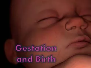
Gestation
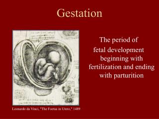
Gestation. The period of fetal development beginning with fertilization and ending with parturition. Leonardo da Vinci, "The Foetus in Utero," 1489. Fertilization. Zygote: diploid cell resulting from the fusion of the male and female pronuclei (syngamy)

Gestation
E N D
Presentation Transcript
Gestation The period of fetal development beginning with fertilization and ending with parturition Leonardo da Vinci, "The Foetus in Utero," 1489
Fertilization • Zygote: diploid cell resulting from the fusion of the male and female pronuclei (syngamy) • Embryo: organism in the early stages of development; not recognizable as a member of a specific species
Cleavage • Following fertilization the zygote undergoes several mitotic divisions inside the zona pellucida (overall size does not change). • 1st cleavage yields a 2 celled embryo, • each cell is called a blastomere and is totipotent • Divisions continue rapidly until the 32 cell stage
Preattachment development of the embryo 8-cell embryo morula early blastocyst hatched blastocyst Senger, pg 286
Maternal Recognition of Pregnancy • Necessary to prevent luteolysis • Occurs prior to implantation/placentation • Differs among species
Pregnancy Recognition Signals • Cow – bovine interferon tau (bIFNt); produced by conceptus, days 15-16 of gestation are critical • Sheep – ovine interferon tau (IFNt); produced by conceptus, days 13-14 of gestation • Sow – Estradiol; produced by conceptus and causes a reroute of PGF2α; must occur days 11-12 • Mare – Protein/estrogen complex and embryo migration; beginning day 12 embyro must migrate 12-15 times per day Senger, pgs 291-295
Differentiation • Formation of three germ layers • Formation of the extraembryonic membranes (placenta) • Formation of organs • Rapid changes in relative size
ICM Blastocoele Three Embryonic Germ Layers • Inner cell mass (ICM) = embryo • Trophoblast = chorion Mesoderm Muscle, skeleton, Cardiovascular system, Reproductive system Ectoderm Nervous system, skin, hair Endoderm Digestive system, lungs, endocrine system Senger, pg 81-82
Extraembryonic Membranes Senger, pg 290
Function of Fetal Membranes and Fluids Yolk Sac – Originates from fetal midgut. Nutrient supply for early embryo. Absorbs uterine sectretions from endometrium to stimulate early embryonic development. Amnion – Innermost membrane directly surrounding the fetus. Protects from injury and provides lubrication for parturition. Prevents lung collapse and opens digestive tract. Allantois – Originates from the gut and forms the umbilicus. Supports blood vessels. Reservoir of nutrients and wastes. Chorioallantois: product of fusion between the allantoic and chorionic membranes Chorion –Outermost membrane in direct contact with uterine tissue. Becomes vascularized by allantoic vessels. Site of hormone production, nutrient and gas exchange.
Placenta PLACENTA: organ of nutrient and waste exchange between the fetal blood and maternal blood PLACENTAL SHAPE: defines the proportion of surface area shared between fetal membranes and maternal uterine tissue where exchange occurs PLACENTAL TYPE: defines the structure of cell layers separating fetal blood from maternal blood
Placental Shapes • Diffuse – noninvasive • Cotyledonary – placentomes are the point of high throughput maternal/fetal contact • Zonary/Discoid – invasive; most direct contact between fetal and maternal blood Senger pgs 308-310
Diffuse Placenta Fetal Chorion Uterine Endometrium Senger, pg 308
Cotyledonary Placenta Senger, pg 310
Cotyledonary Placenta Placentomes Umbilicus Fetus
Types of Placentas Type = degree of invasiveness, based on layers separating maternal blood from the fetal epithelium Least Invasive epitheliochorial synepitheliochorial endotheliochorial Most Invasive hemochorial Maternal Blood Maternal endothelium Maternal connective tissue Maternal epithelium Fetal epithelium Fetal connective tissue Fetal endothelium Fetal Blood
Placentation by Species SPECIESSHAPETYPE Cow Cotyledonary Epitheliochorial Ewe Cotyledonary Synepitheliochorial Sow Diffuse Epitheliochorial Mare Diffuse Epitheliochorial Dog, Cat Zonary Endotheliochorial Primates Discoid Hemochorial Rodents Discoid Hemochorial
Hormones Produced by the Placenta • equine Chorionic Gonadotropin: maintains primary CL, responsible for formation and maintenance of accessory CL. • human Chorionic Gonadotropin: maintain CL • Progesterone: in some species (ewe, mare, woman) the placenta takes over progesterone production later in gestation. ~”progesterone block” – inhibits myometrial contractions • Estrogen: peak of E2 signals preparturient period in some species • Placental Lactogen: stimulates growth of fetus and mammary glands • Relaxin: softens connective tissue in the cervix and relaxes pelvic ligaments
Fetal Growth Growth: period of development from embryo to fully developed fetus, prior to parturition Example of development relative to time for the bovine Calcification of Bone Matrix 70 days Extensive Bone Formation 180 days Tooth Formation 110 days Hair, eyes, muzzle 150 days Hair over entire body 230 days
Relative Development parturition Shift from classification as embryo to fetus ZP Disintegration fertilization placentation Cleavage DifferentiationGrowth More than 50% of the total weight of the fetus at parturition is gained during the last two months of gestation
Parturition The process by which the uterus expels the products of conception
Stages of Parturition • Stage I – preparatory stage involving cervical dilation and positioning of the fetus in the birth canal via myometrial contractions. • Stage II – time of hard labor and expulsion of the fetus. • Stage III – expulsion of the placental membranes and subsequent uterine involution.
Brief Definitions • Dystocia – difficult birth • Usually refers to calving difficulty • i.e., absolute or relative • Presentation – direction of fetal delivery • Anterior, posterior, or transverse • i.e., anterior = head first • Position – orientation of the fetus • Dorsal or ventral side up or lateral • Posture – location of the legs, head, and neck • relates to normal/abnormal posture
Cattle • Dystocia is the major cause of calf loss • Relative Dystocia – normal sized calf and a small birth canal • Absolute Dystocia – abnormally large calf and a normally sized birth canal • Most common cause of dystocia is from an oversized calf • Hiplock – shoulders of the calf “lock” onto the bones of the pelvis during delivery • Results in the calf getting stuck in the birth canal
Cattle • Stages: • Stage I – lasts from 1-4 hours (seldom noticed) • Stage II – begins with the rupture of the allantochorion • Should allow for 2-3 hours for a normal birth to occur without interfering • Stage III – begins after expulsion of the fetus • Lasts up to 4 – 5 hours after fetal expulsion • Presentation: • Anterior • Position: • Dorsal side up • Posture: • Front legs first (slightly offset), bottom of hooves facing down, and nose between front legs
Sheep • The process of parturition in the sheep is very similar to that in cattle • Twins are present if a front and rear leg appear together
Swine • Duration of farrowing (parturition): • 4 – 48 minutes per piglet • Presentation: • Anterior or posterior • Position: • Dorsal or ventral side up • Posture: • Not specific
Horses Foaling time is difficult to predict • Relaxation of the sacrosciatic ligament • Distended mammary glands - days • Waxing – within 48 hours
Foaling • Stages: • Stage I – lasts ~ 1 hour • Restlessness, sweating in flanks • Stage II – very rapid, lasts ~ 10-20 minutes • Stage III – lasts from 1-12 hours
Horses • Presentation: • Anterior • Position: • Dorsal side up • Posture: • Front legs first (slightly offset), bottom of hooves facing down, and nose between front legs • It is crucial that the foal is born within 30 minutes of the beginning of stage II. Failure to do so, will most likely lead to the death of the foal. • Due to the “crushing” of the umbilical cord between the foal and the dam • deprives the foal of oxygen
Hormones of Parturition • ACTH – induces parturition in response to fetal stress • Fetal Cortisol – induces the release of PGF 2alpha and produces the enzymes needed to convert placental progesterone to estradiol • Placental Progesterone – maintains pregnancy and will be converted to estradiol during parturition • Estradiol – enhances secretions of female tract (lubrication) and myometrial contractions, as well as enhance the receptors for PGF 2alpha and oxytocin
Hormones of Parturition • PGF 2alpha – enhances myometrial contractions, induces luteolysis and the release/secretion of relaxin • Relaxin – relaxes the pelvic ligaments, allowing them to stretch for expulsion of the fetus • Oxytocin – enhances myometrial contractions (produces the most forceful contractions)
Induction of Parturition**Not Recommended Unless the Life of the Dam or the Fetus is Threatened** • Inducing parturition can enhance reproductive management by controlling the time of parturition • Dangerous unless parturition is impending (within one day of occurring) • Retained placenta often occurs • Forcing expulsion of the fetus and the fetal membranes (i.e., the placenta) before the placenta is ready to detach from the uterus
Cattle Glucocorticoids Prostaglandins Estrogens Sheep Glucocorticoids If given after day 140, response within 48hrs Swine Glucocorticoids Prostaglandins If given after day 110, response within 30hrs Horses Glucocorticoids Prostaglandins All or none response Agents Used to Induce Parturition Oxytocin: Works well, but the dose and timing are extremely important. All impending signs of parturition must be present. (general rule: give ~ 1hr before parturition)
Neuroendocrine Reflex Arc • Teat stimulation (suckling) • Stimulation of nerves in the spinal cord • Nerve signals to the brain • Release of oxytocin from posterior pituitary into blood • Oxytocin received by the mammary tissue • Contraction of alveoli • Contraction of ducts • Milk ejection 3 4 5 2 5 1 6 7 8
Mammary Gland Anatomy Tissue of Mammary Gland: Secretory cells Alveoli Myoepithelial cells Ducts Blood Vessels
Initiation of Lactation: (lactogenesis) 1. Increased formation and development of duct system 2. Increased formation and development of secretory cells 3. Development of capacity for milk synthesis
Hormones Required for Lactation 1. Estrogen: stimulates duct growth 2. Progesterone: stimulates alveolar development 3. Growth Hormone (somatotropin): stimulates milk production 4. Thyroid Hormones: enhance development and function of mammary tissue 5. Corticoids: enhance synthesis of enzymes necessary for milk biosynthesis 6. Prolactin: initiation of lactogenesis 7. Oxytocin: stimulate myometrial contractions to release milk from alveoli to ducts 8. Insulin: maintain function and survival of secretory cells 9. Placental Lactogen: general mammary tissue growth Adrenalin: prevents milk ejection; blocks secretion of oxytocin, causes vasoconstriction in mammary gland Administration of bST to dairy cattle increases milk production 15-40%. Suggested that bST stimulates insulin-like growth factor-1 (IGF-1) which stimulates the secretory cells.