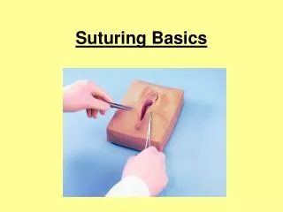
Basic Suturing Skills Workshop
Basic Suturing Skills Workshop. Shirley Pollard Ramsey DNP, FNP-BC, CRNFA Bluegrass Surgical Assistants, PSC Lexington, Kentucky. Ideally wound closure should be effective: simple, efficient, inexpensive, and painless, and should achieve optimal cosmetic results. OBJECTIVES.

Basic Suturing Skills Workshop
E N D
Presentation Transcript
Basic Suturing Skills Workshop • Shirley Pollard Ramsey • DNP, FNP-BC, CRNFA • Bluegrass Surgical Assistants, PSC • Lexington, Kentucky
Ideally wound closure should be effective: simple, efficient, inexpensive, and painless, and should achieve optimal cosmetic results.
OBJECTIVES • Review the Anatomy and Physiology of the Wound Healing Process • Discuss the Guidelines for Seeking Surgical Consultation for Laceration Repair • Assess, Inspect and Evaluate the Patient and Wound • Outline Commonly used Anesthetic Agents, including Pharmacology, onset of action, and duration of action. • Demonstrate administration of local anesthesia for wounds including digit and toe blocks. • Identify the various types and sizes of suture material. • Review and demonstrate basic wound closure techniques including skin lines • Develop a wound care plan and follow-up for suture removal. • Demonstration and Discussion
ANATOMY / SKIN and FASCIA • Epidermis • Dermis • Superficial Fascia • Deep Fascia
THE WOUND HEALING PROCESS • The entire wound healing process is a complex series of events that begins at the moment of injury and can continue for months to years. The wound healing process includes: • I. Hemostasis • II. Inflammatory (preparative) Phase • III. Proliferative Phase (Collagen formation) • IV. Tissue Remodeling (Resolution)Phase
I. Hemostasis • Vascular constriction • Platelet aggregation, degranulation, and fibrin formation (thrombus)
Healing • II. Inflammatory Phase • Inflammation, Vasodilation, Phagocytosis • Neutrophil infiltration • Monocyte infiltration and differentiation to macrophage • Lymphocyte infiltration • Process: 3-7 days
III. Proliferative Phase • Re-epitheliazation • Angiogenesis • Collagen synthesis • Extracellular matrix formation
IV. Remodeling Phase • A) 3 weeks to 2 years • B) New collagen forms which increases tensile strength to wounds • C) Scar tissue is only 80 percent as strong as original tissue Collagen remodeling • Vascular maturation and regression
Guidelines for Seeking Surgical Consultation for Laceration Repair • Deep wounds of the hand or foot • Full-thickness lacerations of the eyelid, lip, or ear • Lacerations involving nerves, arteries, bones, or joints • Penetrating wounds of unknown depth • Severe crush injuries • Severely contaminated wounds requiring drainage • Wounds leading to a strong concern about cosmetic outcome
ASSESS / EVALUATE / PLAN WOUND MANAGEMENT • Note: • Bleeding / Control by direct pressure • History • Mechanism of injury • Age of wound • Personal Health History • Age, human immunodeficiency virus and diabetes status, oxygenation, tobacco use, alcoholism, nutrition, radiation • tetanus immunization history • ALLERGIES to: latex, local anesthesia, tape, or antibiotic • Associated signs and symptoms
ASSESS / EVALUATE / PLAN WOUND MANAGEMENT (cont’d) • Time of incident • Location • Level of Bleeding • Baseline neurovascular and functional status of the involved body part • Involvement of tendons, nerves, blood vessels, or bone involvement should be evaluated before repair • Size and Depth of wound • Shape • Foreign bodies • Contamination
Contradictions to Wound Closure • Redness around edges • Infection • Fever • Edema-excessive • Puncture Wounds • Animal Bites ( MUST BE REPORTED) • Wounds with nerve or tendon damage • Wounds of eye, eye lid, bites (Human & Animal) are very contaminated • Wounds of the thoracic or abdominal cavities • Wound greater than 12 hours
REVIEW Anesthetic Agents and Correct Dosages for the Different Suture Sites • Lidocaine (with or without Epinephrine) • Most commonly used, Rapid Onset • Strength 0.5%,1%,2% • Max Dose 5mg/kg • * Can use with Epinephrine for prolonged effect and decreased bleeding. Dosage is 7mg/kg. • DO NOT USE EPINEPHRINE on Eyes, Nose, Fingers, Toes, Penis, Scrotum, or Ears. • Marcaine (with or without epinephrine) • Slow onset, long duration • Max Dosage 3mg/kg • 0.5% and 1%
INJECTION TECHNIQUES • *** CHECK for ALLERGIES! (True allergies are uncommon, but do occur.) • In persons who are allergic to amides use: 1 ml dyphenhydramine (50 mg/ml) to 4 ml sterile saline for local anesthetic effects • Use 25 gauge needle or smaller to decrease sting of injection • Insert needle at inner edge of wound • Aspirate • Inject anesthetic as you pull the needle out • Buffering with 1 ml of 8.4% sodium bicarbonate (1 ml / 10 ml local anesthetic) to 10 ml of local anesthetic significantly reduces pain of injection
IDENTIFY the various types and sizes of suture material • Absorbable vs. non-absorbable • Natural vs. synthetic • Monofilament vs. multifilament
ABSORBABLE SUTURE • Degraded and eventually eliminated in one of two ways: • Inflammatory reaction utilizing tissue enzymes • Hydrolysis • Examples: • “Catgut” (Untreated Chromic) :Very fragile and non reactive • Chromic ( Catgut treated with chromium salt solution to aid in resisting breakdown): Absorption is 90 days, TSR: 28 days • Vicryl (Polyglactin): TSR: 75% at 2 weeks, 50% at 3 weeks, 25% at 4 weeks • Monocryl: High tensile strength in the first few weeks. TSR: 60 - 70% at 1 week, 30 - 40% at 2 weeks • PDS: Longest retention of the absorbable sutures. 70% at 2 weeks. 50% at 4 weeks. 25% at 6 weeks
NON-ABSORBABLE • Not degraded, permanent • Examples: • Prolene: Does not adhere to tissue (Blepharoplasties) • Nylon: Skin closures • Stainless steel: Now used primarily in sternal closures • Silk* Poor tensile strength if wet • (*not a truly permanent material; known to be broken down over a prolonged period of time—years (1 year plus)
NATURAL SUTURE • Biological origin • Cause intense inflammatory reaction • Examples: • “Catgut” – purified collagen fibers from intestine of healthy sheep or cows • Chromic – coated “catgut”: facial wounds, lip/intraoral mucosa, children’s wounds. Lasts 7-14 days at most. • Silk
SYNTHETIC SUTURE • Synthetic polymers • Do not cause intense inflammatory reaction • Examples: • Vicryl / SH • Monocryl / SH / PS-2 • PDS • Prolene • Nylon FS-2
MONOFILAMENT SUTURE • Grossly appears as single strand of suture material; all fibers run parallel • Minimal tissue trauma • Resists harboring microorganisms • Ties smoothly • Requires more knots than multifilament suture • Possesses memory • Examples: • Monocryl, PDS, Prolene, Nylon
MULTIFILAMENT SUTURE • Fibers are twisted or braided together • Greater resistance in tissue • Provides good handling and ease of tying • Fewer knots required • Examples: • Vicryl (braided) • Chromic (twisted) • Silk (braided)
SUTURE SIZES • Sized according to diameter with “0” as reference size • Numbers alone indicate progressively larger sutures (“1”, “2”, etc) • “1”: VERY LARGE, USED TO CLOSE THE ABDOMINAL WALL • Numbers followed by a “0” indicate progressively smaller sutures (“2-0”, “4-0”, etc) • “10-0”: very tiny (fine as a human hair, used for microvascular anastomoses and corneal sutures) • COMMON SIZES: 2-0 to 5-0
NEEDLE TYPES • CURVED • Designed to be held with a needle holder • Used for most suturing • CUTTING: Used primarily for suturing skin. • TAPERED: Used to suture soft tissue, excluding skin (e.g. GI tract, muscle, fascia, peritoneum) • STRAIGHT • Often hand held • Used to secure percutaneously placed devices (e.g. central and arterial lines) • FS , PC, PS, SH, RV, UR,
NEEDLES • Cutting Needle • Triangular body • Sharp edge toward inner circumference • Used to suture skin or tough tissue • Taper-point Needle • Round body • Used to suture soft tissue, excluding skin (e.g. GI tract, muscle, fascia, peritoneum)
Wound Closure • Primary • Secondary • Tertiary
Primary Closure • Primary Closure • Face or Scalp: Repair within 24 hours (18 hours preferred) • Body: Repair within 12-18 hours (6 hours preferred)
Secondary Closure(Older Wounds with Infection Risk) • Step 1: Initial Evaluation • Option 1: Loose approximation with simple interrupted closure • Option 2: Pack wound with sterile wet to dry dressings changed twice daily • Step 2: Reevaluation at 3-5 days • No infection: Primary wound closure • Infection: Treat infection and healing by second intention as below • Healing by second intention • Pack wounds with sterile wet to dry dressing bid • Granulation and contraction risk without suturing
Tertiary Closure • Two surfaces of granulation tissue are brought together after debridement of nonviable tissues and left open • Contaminated, dirty and infected traumatic wounds with extensive tissue loss and a high risk of infection • wound gradually gains sufficient resistance to infection which permits an uncomplicated closure • takes place 4 to 6 days postinjury.
REVIEW SKIN LINES • Two types of skin tension lines • Important impact on final healing • Static (Langer’s Lines) skin is under constant tension • Dynamic (Kraissl’s Lines) forces created by muscles and correspond to wrinkles • Parallel repairs are less likely to scar • Repairs under minimum tension are less likely to scar
KNOT TYING PRINCIPLES • Simple knots are best • Smaller knots are better than big ones • Use the minimum ties per knot • Tension should be as horizontal as possible (don’t lift) • Excessive tension will cause tissue damage • The two-handed square knot is the easiest and most reliable knot for tying most suture material
REVIEW SIMPLE SKIN CLOSURES • Start on the side of the wound opposite and farthest from you to ensure that you are always sewing toward yourself. By sewing toward yourself, the suturing process is made easier from a biomechanical standpoint. • 1. Start from the outside of the skin, go through the epidermis into the subcutaneous tissue from one side, then enter the subcutaneous tissue on the opposite side, and come out the epidermis above. • 2. To evert the edges, the needle tip should enter at a 90° angle to the skin. Then turn your wrist to get the needle through the tissues. • 3. You can use simple sutures for a continuous or interrupted closure. • The needle tip should enter the tissues perpendicular to the skin. Once the needle tip has penetrated through the top layers of the skin, twist your wrist so that the needle passes through the subcutaneous tissue and then comes out into the wound. This technique helps to ensure that skin edges will evert. • The SIMPLE INTERRUPTED is the technique of choice if you are worried about the cleanliness of the wound. • If the wound looks like it is becoming infected, a few sutures can be removed easily without disrupting the entire closure. • Interrupted sutures can be used in all areas but may take longer to place than a continuous suture.
DEMONSTRATE ADVANCED SKIN CLOSURES • VERTICAL MATTRESS SUTURE • HORIZONTAL MATTRESS SUTURE • RUNNING SUBCUTICULAR SUTURE
VERTICAL MATTRESS SUTURE • Affords precise approximation of skin edges with eversion • Two-step stitch: • Simple stitch made – “far, far” relative to wound edge (large bite) • Needle reversed and 2nd simple stitch made inside first – “near, near” (small bite)
HORIZONTAL MATTRESS SUTURE • Provides hemostasis and added strength in fascial closure; also used in calloused skin (back, palms, soles) • Two-step stitch: • Simple stitch made • Needle reversed and 2nd simple stitch made adjAcent to first (same size bite as first stitch)