Download
1 / 66
720 likes | 956 Vues
The Pathology of Viral Infections. Doç.Dr.A.Işın DOĞAN EKİCİ Department of Pathology. The Virus es & Virus induced cell injury.

E N D
ThePathology of Viral Infections Doç.Dr.A.Işın DOĞAN EKİCİ Department of Pathology
The Viruses & Virus induced cell injury • Cytolytic viruses destroy cells, while cytopathic viruses alter their morphology without causing necrosis. Viruses may kill a cell by scrambling the genes it needs to maintain its structure, or by multiplying so fast that the cell explodes. • Most interesting are the "giant cells" which result when several virus-infected cells fuse. • Viral inclusion bodies are crystalloids of viruses in the nucleus and/or cytoplasm. • Further, the body's T-cells often attack cells which harbor viruses. • (e.g, the cell damage in hepatitis B is brought about by T-cells, not viruses.)
Viruses are common cause of human illness, yet most human viral infections are asymptomatic and unrecognized. • Different viruses can produce the same pathologic features (e.g., upper respiratory tract infections), and the same virus (e.g., cytomegalovirus) can produce different diseases according to the host’s resistance and age. • Viruses are obligate intracellular parasites, and are able to commandeer eukaryotic cells to make copies of themselves. • The infectious particle itself is called a virion. The protein-based coat is the capsid, which surrounds nucleic acid (DNA or RNA, never both).
Each virus must (1) attach to the cell, (2) penetrate it, (3) un-coat, and (4) replicate. These stages together constitute the virus cycle. • Viremiameans viruses in the bloodstream. Except for some respiratory viruses, all viruses probably travel via the blood.
Double-stranded DNA viruses (DNA viruses replicate in the nucleus) • Adenovirus family • Hepadnavirus family • Hepatitis B • Herpes viruses • Cytomegalovirus (CMV, HHV 5) • Epstein-Barr virus (EBV, HHV 4) • Herpes simplex 1 • Herpes simplex 2 • Human herpes virus 6,7 (HHV 6,7) • Human herpes virus 8 (HHV 8, Kaposi’s sarcoma) • Varicella/Herpes zoster (VZV, HHV 3) • Papovavirus family • Human papillomavirus • JC virus (PML, brain disease) • Poxvirus family • Molluscum contagiosum • Smallpox • Smallpox vaccine (vaccinia)
RNA viruses(RNA viruses replicate in the cytoplasm) • Reovirus family • Rotavirus (sporadic viral gastroenteritis) • Coronavirus family • Orthomyxovirus family • Influenza group • Picornavirus family • Calicivirus subfamily Hepatitis EWinter vomiting viruses • Enterovirus subfamily • Coxsackie virus • Echovirus • Poliovirus • Many others • Hepatitis A • Hand-foot-mouth disease • Rhinovirus subfamily
Paramyxovirus family • Measles • Mumps • Parainfluenza • Respiratory syncytial virus • Metapneumovirus • Retrovirus family • HIV-1 & 2 • HTLV I & II • Animal tumor viruses • Togavirus family • Rubella • Hepatitis C • Hepatitis G
Arboviruses (toga-, flavi-, arena-, bunya-, reo-, filo-) • Arbovirus encephalitis viruses • Colorado tick fever • Dengue family • Regional hemorrhagic fevers • Crimien Congo hemorrhagic fever • Yellow fever • Hantavirus • West Nile Virus • Other • Parvovirus • Rabies virus
Depending on their type, viruses cause hazard by: (1)destroying host’s cells as their progeny are released (lytic infection, typical of herpes viruses, smallpox); (2)rendering infected cells non-functional (HIV); (3)exciting cell-mediated immunity, which destroys otherwise-healthy cells which happen to be infected by the virus (hepatitis B, infectious mononucleosis). (4)causing cell overgrowth, which may be unsightly (warts, molluscum), a fertile ground of carcinogenesis (Epstein-Barr virus in Africa), or full-blown malignancy (some human papillomavirus infections). (5)some viruses promote cell fusion (measles and herpes produce picturesque giant cell formation; the nuclei, loaded with visible virus aggregates; multinucleated brain microglial cells are markers for AIDS).
Virus invasion and Host immune responses Viral entry can be via: • respiratory, • gastrointestinal, • genitourinary tract, or • it can bypass the powerful skin or mucosalbarriers by insect bite (arboviruses), • animal bite (rabiesvirus), • wound or minor abrasion (HIV), • needle (Hepatitis B, HIV). To induce a systemic disease, a virus must successfully overcome immune defenses and must: - attach to nearby cells, - replicate within them, - spread from the entry site to distant target cells (via bloodstream, lymphatics, and nerves).
Viral inclusions are aggregates of virus proteins, visible by light microscopy: 1. Intranuclear (Cowdry A and Cowdry B) • Adenovirus (smudge cells) • Cytomegalovirus (one large, clearly-defined) • Herpes simplex 1&2 (one large, clear, + multinucleate) • Herpes zoster (one large, clear, + multinucleate) • Measles (Warthin-Finkeldey cells) 2. Intracytoplasmic • Cytomegalovirus (many small) • Rabies (Negri bodies in neurons) • Molluscum contagiosum (molluscum bodies in skin) • Smallpox (Guarnieri bodies in skin).
Herpes simplex-Cowdry A Measles (Warthin-Finkeldey cells)
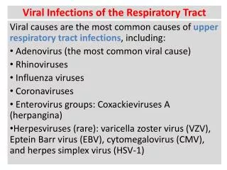
RESPIRATORY VIRAL DISORDERS • These contagious diseases, typically spread by droplets, range from "the common cold" to mortal viral pneumonitis. • Upper respiratory infections involve the nose, sinuses, throat, tonsils, and/or middle ear. • The anatomic distribution is variable, and sinuses are often occluded. • Lower respiratory infections involve the larynx, trachea, bronchi, alveoli (viral pneumonitis, chest cold), and/or pleura. • Viral infection of the respiratory tract and lungs predispose to bacterial superinfection.
The PATHOLOGY of Upper respiratory tract infections Mucosal hyperemia with abundant mucus production and edema (serous exudate), and a preponderance of lymphocytes, monocytes and plasma cells. • The swollen mucosa and viscid exudate may plug the nasal channels, sinuses, or eustachian tubes and lead to suppurative secondary bacterial infection.
Etiology: • Common cold • Picornaviruses, especially rhinoviruses andcertain echoviruses and Coxsackieviruses • Grippe; Grip; Flu • Influenzavirus • Many respiratory illnesses varying from the common cold to influenza-like pneumonia • Parainfluenzavirus
Other viral respiratory tract infections: • Virus induced tonsillitis • Laryngotracheobronchitis and bronchiolitis • Viral obliterative bronchiolitis • SARS • Viral pneumonia
SEVERE ACUTE RESPIRATORY SYNDROME(SARS) • (SARS) is a viral respiratory tract infection first detected in the Guangdong province of China in late 2002. It has since spread to at least 17 countries, mainly in Southeast Asia, but also to Canada and the United States. • SARS appears to be caused by a strain of coronavirus. • Transmission occurs to close personal contacts (eg, health care workers; family members; and persons in nearby seats on airplanes, beds in hospitals, and rooms in hotels), leading to presumption of respiratory droplet spread.
Eighty to 90% of patients have a mild illness and recover within 1 to 2 weeks. • The remaining 10 to 20% develop respiratory distress with marked dyspnea, hypoxemia, and occasionally adult respiratory distress syndrome (ARDS).
Viral pneumonia • Interstitial inflammatory reaction (edema, and mononuclear infiltration) fill the alveolar septa, but without entering the alveolar spaces. • In severe cases, type II pneumocytes round up and desquamate into the alveolar spaces with the formation of hyaline membranes layering the septal walls. • In severe, fulminating, influenzal viral pneumonia, widespread fibrinohemorrhagic alveolar consolidation is superimposed. • Adenovirus and herpesvirus families have the power to necrosis, and evoke neutrophilic exudation.
Systemic complaints begin 1-2 days after exposure, with fever, headache, myalgias, and fatigue. • In severe cases, staphylococcal superinfection is common and deadly. • Tens of thousands of people die of influenza during every epidemic. • Note that certain other viruses (measles, mumps, rubella, chickenpox, etc.) enter the body by way of the lungs. • They may produce lung involvement during the clinical phase, but this follows spread of the virus through the bloodstream.
VIRAL DISORDERS of DIGESTIVE TRACT Mumps Mumps is an acute, contagious childhood (in 5- to 15-year-old) disease featuring transient inflammation and swelling of the major salivary glands. Asymptomatic or very mild infection is common. The parotitis is bilateral in about 70% of cases or unilateral in 20%, but in about 10% there is only sublingual involvement. Affected glands are enlarged and painful.
Most but not all adults are immune. • Adults may also suffer from orchitis, oophoritis, and/or pancreatitis. • Testicular swelling may contain parenchymal hemorrhage. • High-pressure in the testis is uncomfortable and is likely to cause ischemia and permanent loss of spermatogenesis=infertility! • Fortunately, it is usually unilateral.
Microscopically, the gland interstitium is edematous and diffusely infiltrated by histiocytes, lymphocytes, and plasma cells, which compress acini and ducts. • In the pancreas, lesions become more destructive, causing parenchymal and fat necrosis and polymorphonuclear cell infiltration. • Similarly, in the testis, tightly contained by the tunica albuginea, tissue swelling may result in necrosis of seminiferous tubules with neutrophilic infiltration and focal hemorrhages and micro infarctions, and finally scars (mumps orchitis). • Children should be immunized against mumps.
Viral enteritis & diarrhea • Viruses are major causes of acute diarrheal diseases previously attributed to uncertain causes. • Rotavirus is an important cause of mild winter vomiting and diarrhea in children; most adults are immune. • Norwalk agent causes outbreaks of vomiting and/or diarrhea at any time, in children or adults. • Bowel biopsies have shown mixed inflammatory infiltration of the lamina propria, shortening of villi, and cellular hyperplasia of mucosal crypts.
VIRAL DISORDERS with EXANTHEMS and SKIN RASHES • First disease: measles (rubeola) (Kızamık) • Second disease: scarlet fever (kızıl) • Third disease: German measles (rubella) (Kızamıkçık) • Fourth disease: Filatov-Dukes' disease • Fifth disease: erythema infectiosum (slapped cheek syndrome) (Parvo Virus) • Sixth disease: roseola infantum (herpesvirus 6 infection; exanthem subitum)
Measles (Rubeola) “Kızamık” • Measles is a major infectious disease among children 3 to 7 years old. • Before the vaccine, measles was part of everyone's childhood. • Transmission is by droplets. The incubation period is two weeks. Photophobia and eye-burning are the first symptoms. • It is an acute, febrile, systemic viral infection usually beginning with • coryza and conjunctivitis, • followed by typical spotty lesions inside the mouth (Koplik's spots; blister-ulcers next to Stensen's ducts), • lymphoreticular hyperplasia, • and a blotchy, generalized, erythematous rash. Koplik's spots are diagnostic even before the rash begins.
Histologically, the rash is produced by dilated skin vessels, edema, and a moderate, nonspecific, mononuclear perivascular infiltrate. • Ulcerated mucosal lesions in the oral mucosa (Koplik's spots) are marked by necrosis, neutrophils, and neovasculazition. • The lymphoid organs typically have marked follicular hyperplasia, large germinal centers, and randomly distributed multinucleate giant cells (Warthin-Finkeldey cells), with eosinophilic nuclear and cytoplasmic inclusion bodies.
HERPESVIRUS DISEASES • These are important DNA viruses. • Each usually produces a mild (often unnoticed) illness upon entering the body. • The virus then lies latent within the host genome, awaiting reactivation, for the rest of the person's life; and from time to time, herpes infections kill people. • Patients treated for lymphomas and recipients of bone marrow transplants are notoriously susceptible.
The spectrum of HSV I and HSV II infections includes the following: 1.Latency (the virus slowly reproduces in ganglia, trigeminal or other; diagnosis is made only by antibody titer) 2.Skin or mucosal vesicles (fever blisters, cold sores, gingivostomatitis, genital herpes) 3.Severe vesicular eruption of the eye or skin (herpes keratoconjunctivitis, eczema herpeticum, Kaposi’s varicelliform eruption) 4.Severe CNS lesions (herpes simplex encephalitis, transverse myelitis) 5.Opportunistic localized lesions in internal organs (herpes esophagitis, herpes simplex pneumonitis) 6.Overwhelming disseminated opportunistic infection (with focal necrosis of many organs in neonates and compromised adults).
Chickenpox (varicella) & Herpes zoster Chickenpox • On first meeting this virus, a child develops chickenpox, a highly contagious disease spread by droplets. • The rash consists of vesicles which arise in successive crops over the body (starts on the trunk and spreads outward, extremities and face, centrifugally). • Blistering may also occur on the buccal mucosa. Severe form may resemble hemorrhagic smallpox (focal hemorrhages within or around the blisters). • The condition resolves within about a week. • Older patients can get an encephalitis or pneumonitis which is probably immune-mediated.
By electron microscopy, viral particles of any herpesvirus appear as arrays and scattered single particles as shown here in a nucleus of a neuron from the cerebrum from a patient with herpes simplex encephalitis.
Severe neonatal disease may occur when the mother becomes infected during the first 20 weeks of pregnancy, but unlike German measles, infection during gestation rarely induces congenital malformations in the offspring. • A varicella virus vaccine is available in general use. • After recovery, the virus hides out in the dorsal root and/or sensory trigeminal ganglia.
Herpes zoster (zoster, shingles) • It is recurrence of the chickenpox rash, typically along the distribution of a sensory nerve root. • During the attack of zoster, reactivated virus travels centrifugally from the ganglia to the skin of the corresponding dermatomes, resulting in a localized vesicular eruption that is similar to that of chickenpox • but differentiated by the often intense itching, burning, or sharp pain in the affected skin segment because of a simultaneous radiculoneuritis. • It is preceded by paresthesias, and pain after herpes zoster is notorious (post-herpetic neuralgia). • Involvement of geniculate ganglion may induce a facial paralysis (Ramsay Hunt syndrome).
Herpes simplex 1 • Herpes simplex is transmitted by physical contact such as kissing, and it thus spread among family members and friends. • A majority of adults have the virus, though most never become sick or suffer only fever blisters. • About half of all babies are born with IgG antibodies to this agent transmitted across the placenta. • The virus climbs sensory nerves and hides in nervous tissue (especially trigeminal ganglion). • This is typically a necrotic area on the lower lip, also called a cold sore, fever blister, sun blister, stress blister.
Herpesvirus lesions, wherever located, are marked by cytopathic changes, principally the formation of Cowdry type A intranuclear inclusions • However, cell fusions may produce inclusion-bearing polykaryons or giant cells, which can also be found in smears of blister fluid (Tzanck preparations), confirming the diagnosis of a herpetic infection.
Herpes simplex II(Herpes genitalis) • This virus is usually contracted through sexual contact, and produces painful, recurrent blisters on the genitals. • The pathology is identical to HSV-I. • In the immunocompromised, HSV-2 may be more harmful. • If active herpes simplex is present in the birth canal, the newborn will probably contract the infection during vaginal delivery. This is often a fulminant, fatal infection. When in doubt, Caesarean section is indicated.
Cytomegalic inclusion disease(CID) A very common viral infection caused by the cytomegalovirus (CMV; HHV 5), a member of the herpesvirus group. It can be contracted by; 1.intrauterine transmission of a latent infection in the mother, 2.perinatal transmission to the fetus as it passes through the birth canal of a mother with a cervicovaginal infection, 3.respiratory droplet transmission among children and possibly between adults, 4.blood transfusions (Pope John Paul II got sick from transfusion), 5.transplantation of virus infected grafts (especially bone marrow), 6.veneral transmission via semen or vaginal fluid, 7.transmission through mother’s milk.
Teens and adults may note an infectious mononucleosis-like infection which lasts up to a few weeks. • Despite the high frequency of infection in adults, it is almost always latent and asymptomatic unless the patient is immunocompromised (e.g., AIDS). • The most disturbing feature of CMV is its ability to damage the unborn child. • While most fetuses meeting CMV show no signs of damage, a child with congenital CMV syndrome is small for gestational age,hepatosplenomegaly (due to extramedullary hematopoiesis), jaundiced, afflicted with hemolytic anemia, afflicted with thrombocytopenia with purpurae and pneumonia, blind, deaf, retarded, and/or epileptic (closely resembles erythroblastosis fetalis).
Findings are depend on the severity of the Infection in neonatal disease: - lungs (edema, proteinaceous exudate, focal hyaline membranes), - intestines (focal necroses, punch-out ulcerations), - brain (focal acute inflammation, irregularly scattered necrotic lesions typically occurring around the ventricles and calcifying, microcephaly or hydrocephalus).
The hallmark of any CMV infection is very large cells with large pleomorphic nuclei harboring intranuclear inclusions. • The inclusion is huge and is surrounded by a clear halo. • There are also small basophilic inclusions in the cytoplasm. • The best single place to look inclusions is salivary glands. • Next are kidneys, lungs, and the endocrine glands. Cytomegalic inclusions are often abundant in the renal tubular epithelium. • Cells so affected may not induce inflammatory reactions, but others die and evoke a leukocytic response.
CID much depends on the immunologic competence of the adult. • In compromised host (e.g., AIDS), CMV may produce an opportunistic infection, with involvement of organs except brain: - lungs (edema, proteinaceous exudate, focal hyaline membranes pneumonia), - intestines (ulcerations perforations) - eye (chorioretinitis blindness).
Infectious mononucleosis (Epstein-Barr virus; EBV; HHV 4) • Infectious mononucleosis is a benign, self-limited lymphoproliferative disease caused by EBV, one of the herpesviruses. • Most people meet the Epstein-Barr virus in childhood and never become symptomatic. • Teens and adults are prone to get the familiar infectious mononucleosus syndrome. • The virus is transmitted by saliva (i.e., kissing). • The incubation period for infectious mononucleosis is about 6 weeks.
The virus has a special trophism for B-cells, in which it multiplies. Eventually, B-cells that bear viral antigens are eliminated by cell-mediated and humoral immunity. • The patient with infectious mononucleosis has fever, malaise, fatigue (interleukin effect), and generalized lymphadenopathy (behind the ears). Usually, the throat is sore. • Around the half of patients get cold agglutinins and autoimmune hemolytic anemia. There is a mild thrombocytopenia. • Proliferation of B-cells bearing EBV antigens on their surfaces is upsetting to T-suppressor cells and T-killer cells. These T-cells (not monocytes) proliferate throughout the body, and are seen in the bloodstream.
Morphology in IM • The peripheral blood (smear): • - Absolute lymphocytosis (total white cell counts between 12000-18000, 95% of which are lymphocytes), • - These are atypical lymphocytes or virocytes, and can be recognized by their large size, abundant vacuolated and basophilic cytoplasm, and large, reticulated nuclei.