Download
1 / 50
590 likes | 1.06k Vues
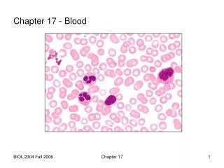
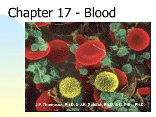
Chapter 17 - Blood. J.F. Thompson, Ph.D. & J.R. Schiller, Ph.D. & G. Pitts, Ph.D. Chapter 17 - Blood. Use the video clips to review blood cell morphology CH 17 RBC Morphology CH 17 WBC Morphology. Overview: Composition of Blood. A liquid connective tissue A mixture

E N D
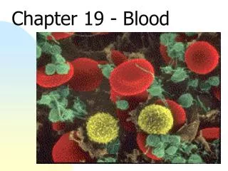
Chapter 17 - Blood J.F. Thompson, Ph.D. & J.R. Schiller, Ph.D. & G. Pitts, Ph.D.
Chapter 17 - Blood • Use the video clips to review blood cell morphology • CH 17 RBC Morphology • CH 17 WBC Morphology
Overview: Composition of Blood • A liquid connective tissue • A mixture • the formed elements - living blood cells & platelets • the plasma – the fluid matrix • Denser and more viscous than water • due to dissolved ions & organic molecules, especially plasma proteins, and to the blood cells • composition and volume regulated by CNS & hormones • Temp - 38°C • pH - 7.4 (critical to be between 7.35 and 7.45) • Volumes differ between sexes, conditional on many factors • Females - average 4-5 L • Males - average 5-6 L
Functions of Blood • Transport and Distribution • delivery of O2, nutrients, and hormones • removal of CO2 and metabolic wastes • Regulation of Internal Homeostasis • body temperature • pH • fluid volume • composition of the interstitial fluid/lymph • Protection • necessary for inflammation and repair • prevents blood loss by hemostasis (coagulation) • prevents infection
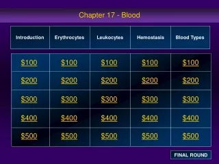
Overview: Composition of Blood • Blood sample • spin it • separates into 2 parts • plasma • ~55% of the volume • straw colored liquid on top • formed elements - ~45% of the volume • red blood cells • buffy coat - white blood cells and platelets • Hematocrit = “packed cell volume” • percentage of formed element measured in a blood sample • about 45%
Blood Components Refer to Tables 17-1 and 17-2 In your text.
Components of Blood - Plasma • Plasma • 92% water • 7% proteins • 1% other solutes
Components of Blood - Plasma • Proteins important for osmotic balance • albumin (60%) • transports lipids • steroid hormones • fibrinogen (4%) - blood clotting • globulins (35%) – many different proteins with a wide variety of functions • globulin classes α, β, and γ • 1% other regulatory proteins
Components of Blood - Plasma • Other solutes • Waste products - carried to various organs for removal • Nutrients – glucose and other sugars, amino acids, lipids, vitamins and minerals • Electrolytes (ions) • Regulatory substances • enzymes • hormones • Gases - O2, CO2, N2
Components of Blood - Formed Elements • Formed elements • >99% red blood cells • <1% white blood cells and thrombocytes (platelets)
Components of Blood - Formed Elements • Erythrocytes, or Red Blood Cells (RBC’s), for O2 and CO2 transport • RBCs’ hemoglobin also helps buffer the blood IMPORTANT! Note the differences in relative size and appearance!
Components of Blood - Formed Elements • Leukocytes (White Blood Cells) • Granular leukocytes (granulocytes) • neutrophils • eosinophils • basophils • Agranular leukocytes (agranulocytes) • lymphocytes - T cells, B cells • monocytes tissue macrophages • Thrombocytes (platelets)
Hematopoiesis • Blood cell formation • All blood cells come from pluripotent hematopoietic stem cells (hemocytoblasts) • reside in red bone marrow • give rise to 5 types of precursor cells • precursors develop into RBCs, WBCs and “giant” megakaryocytes which produce platelets by cytoplasmic fragmentation
Production of Erythrocytes • Erythropoiesis • RBC production • controlled by hormones, especially erythropoietin (EPO) from the kidney • three phases of RBC maturation • production of ribosomes • synthesis of hemoglobin • ejection of the nucleus and reduction in organelles • leave bone marrow as reticulocytes mature in the blood stream to become erythrocytes
RBC Production - Erythropoiesis (cont.) • Reticulocyte count • Reticulocyte • final stage before mature RBC • released into blood where final maturation occurs • Count reticulocytes to evaluate the health of the marrow stem cells or the response of red bone marrow to erythropoietin (EPO) • low count - bone marrow not responding • high count - replacement production or abnormal circumstances
Production of Erythrocytes • Regulation of RBC production • regulated by negative feedback • O2 levels monitored in kidneys • hypoxia increases RBC production • production stimulated by erythropoietin (EPO) from kidneys • Numbers • ♂ - 5.4 million RBC’s/ml (testosterone stimulates EPO synthesis) • ♀ - 4.8 million RBC's/ml • 2 million cells released into blood/second
RBC Production - Anemia • Anemia – symptoms of reduced O2 carrying capacity of the blood • Causes • Insufficient number of RBC’s • hemorrhage - loss of RBC’s • hemolytic anemia - premature RBC destruction due to transfusion reaction, various diseases, or genetic problems • aplastic anemia • destruction or inhibition of hematopoietic components in bone marrow • tumors, toxins, drugs, or irradiation • Decreased hemoglobin content in the RBCs • iron (heme) deficiency - insufficient iron due to diet or poor absorption • pernicious anemia - lack of Vitamin B12 • Vitamin B12 • common in the diet • needed for developing RBC cell division • intrinsic factor needed for proper B12 absorption, often deficient and the actual cause of the B12 deficiency
RBC Production - Anemia • Abnormal Hgb - hereditary • Thalassemias • Greeks, Italians (Mediterraneans) • reduced or absent globin synthesis • RBC’s delicate - may rupture • low RBC count • Sickle Cell Anemia • Africans, African-Americans • Substitution mutation of 1 AA in the hemoglobin molecule changes the shape, flexibility & lifespan of the RBCs • prevents adequate O2 transport • sickled cells lodge in and block capillaries • Need two copies of the abnormal recessive gene for Sickle Cell Disease • One normal, one abnormal copy: increased resistance to malaria = Sickle Cell Trait sickled cells
RBC Production - Erythropoiesis (cont.) • Hematocrit (Hct) • % of blood that is RBC’s • ♂: 40-54% (47%), ♀: 38-46% (42%), Why? • Indicates RBC production and state of hydration • Abnormal Hct • high altitude – hypoxia • athletes - blood doping • polycythemia • anemias • hemorrhage • malaria • cancer • chemotherapy • radiation • drugs
RBC Structure • ~280 million Hgb molecules/cell • Hgb for O2 transport • Bi-Concave shape • greater surface area/volume ratio increases gas diffusion • flexibility allows passage through narrow capillaries
RBC Physiology • O2 combines with Hgb in lungs • O2 gas not very soluble in H2O • Hemoglobin transports O2 Hemoglobin • 2 αglobin chains & 2 βglobin chains • 4 heme groups (lipid) • each heme binds an iron ion (Fe²+) that carries 1 O2
Eosinophil 2-4% Lymphocyte 20-25% Monocyte 3-8% Neutrophil 60-70% Basophil 0.5-1% Differential WBC Count
Lymphocytes - Physiology • Immune response through lymphocytes responding to antigen (AG) • An antigen is: • any chemical substance recognized as foreign when introduced into the body • substance (usually proteins) that stimulate immune responses
Lymphocytes - Physiology • Two main types of lymphocytes • B-cells • particularly active in attacking bacteria • develop into plasma cells to produce antibodies (Ab) • bind to antigen to form antibody-antigen (Ag-Ab) complexes • complexes prevents Ag from interacting with other body cells or molecules • memory B cells – dormant until future exposure to Ag • T-cells • attack viruses, fungi, transplants, cancer, some bacteria • 4 types of cells • cytotoxic (killer) T cells - destroy foreign invaders • helper T cells - assist B cells and cytotoxic T cells • suppressor T cells – help bring immune response to an end • memory T cells - dormant until future exposure to Ag
Leukocyte Life Span and Number • Life span determined by activity • Ingesting foreign organisms, toxins, shortens life • Healthy WBC's – majority last days, but some last months to years • During infection, WBCs may only live hours • engorge with ingested organisms, necrotic cells, toxins, Ab-Ag complexes • often die and lyse (burst)
Leukocyte Life Span and Number • 5,000 - 10,000 WBC’s/mm3 blood • RBC/WBC ratio 700/1 • Differential WBC count (a standard clinical lab report) • Neutrophils 60-70% • Lymphocytes 20-25% • Monocytes 3-8% • Eosinophils 2-4% • Basophils 0.5-1% • Abnormal proportions are correlated with different types of disease processes
Leukocyte Number Abnormalities • Leukopenia = decreased numbers • malnutrition, chronic disease states • drug induced - glucocorticoids, anti-cancer drugs, etc. • Leukocytosis = increased numbers • Normal component of inflammatory response to injuries and infections • Leukemia, Lymphomas = grossly increased numbers, abnormal forms; many subcategories • bone marrow and blood stream (leukemia) or tissue spaces (lymphoma) fill with cancerous (nonfunctional) leukocytes • crowds out other cells types • anemia • bleeding • immunodeficiency
Leukocyte Disorders • Generally a descendent of a single cell • different types of cells • myelocytic leukemia • lymphocytic leukemia • under different cancerous conditions • acute - if derived from -blast type cells • chronic - if derived from later stages
Thrombocytes - Platelets • Development • Megakaryocytes shed small cytoplasmic fragments • Each fragment surrounded by plasma membrane • Anatomy • 250,000-400,000/mm3 • No nucleus, disc shaped • 2-4 µm diameter with many granules
Thrombocytes - Platelets (cont.) Physiology • Short life span (5-9 days) • Help plug small holes in blood vessels • Granules contain regulatory factors which serve several important functions in: • coagulation • inflammation • immune defenses
Thrombocytes - Platelets (Granules) • alpha granules • clotting factors • platelet derived growth factor (PDGF) • dense granules • Ca++, ADP, ATP • Thromboxane A2, • vasoconstrictors • clot promoting enzymes
Hemostasis • 3 mechanisms exist to stop bleeding • First - Vascular Spasm • Blood vessel constricts when damaged • vessel wall smooth muscle contracts immediately • blood flow slows through vessel • local trigger or autonomic reflex?
Hemostasis (cont.) • Second - Platelet Plug Formation • Platelet adhesion • platelets stick to exposed collagen • tissue factors activate platelets • Platelet release reaction • platelets attach to other platelets • release granule contents (thromboxane A2) • promote vasoconstriction, platelet activation and aggregation • Platelet aggregation platelet plug • blocks blood loss in small vessels • less effective in larger vessels
Hemostasis (cont.) • Third - Coagulation • Gel formation (clotting) in blood plasma traps the formed elements • Thrombosis - clotting in a normal vessel • Hemorrhage - slowed clotting may lead to bleeding
Hemostasis - Coagulation • A complicated process that functions as a positive feedback cascade • Fibrinogen Fibrin traps blood cells • 2 pathways – extrinsic & intrinsic unite in a common final process • Pathways involve 12 numbered factors and additional helpers (esp. Ca++) in clot formation
Hemostasis - Coagulation (cont.) • Stage 1: Prothrombinase formation • Prothrombinase catalyzes Prothrombin conversion to Thrombin • Stage 1 has 2 parts • Part 1: Extrinsic Pathway • Rapid (seconds) • Tissue factor (TF) enters blood from tissue • Ultimately activates prothrombinase Extrinsic Pathway Intrinsic Pathway Prothrombinase
Hemostasis - Coagulation (cont.) • Stage 1: Prothrombinase formation (cont.) • Part 2: Intrinsic Pathway • Slower (minutes) • Activators in blood – from damaged red blood or endothelial cells activate clotting • Extrinsic pathway also activates Intrinsic pathway • Ultimately activates prothrombinase • Ca2+ is required for activation of both paths! Extrinsic Pathway Intrinsic Pathway Prothrombinase
Hemostasis - Coagulation (cont.) • Stage 2 - Common Pathway • Thrombin Formation • requires enzyme Prothrombinase & Ca++ ions • catalyzes prothrombin thrombin • Thrombin accelerates formation of prothrombinase (positive feedback) • Thrombin accelerates platelet activation (positive feedback) + + Prothrombinase 2. Common Pathway
Hemostasis - Coagulation (cont.) • Stage 3 - Common Pathway • Fibrin formation • activated enzyme thrombin with Ca++ ions catalyzes fibrinogen fibrin • fibrinogen (soluble) • fibrin (insoluble) • Fibrin • Protein threads attach to vessel and tissue surfaces • Absorbs & inactivates 90% of thrombin, limits clot formation 3.
Hemostasis - Coagulation (cont.) • Clot retraction and repair • clot retraction is also known as syneresis • platelets continue to pull on fibrin threads closing wound • formed elements are trapped in fibrin threads, some serum may leak out • Hemostatic control mechanisms • important that clot formation remains local, not systemic • several mechanisms work together: • fibrin absorbs remaining local thrombin • removal of local clotting factors - washed away • endothelial cells inhibit platelet aggregation
Hemostasis - Coagulation (cont.) • Fibrinolysis - dissolution of a clot, begins within 2 days • plasminogen trapped in clot • many factors convert plasminogen into plasmin (fibrinolysin) • thrombin • activated factor XII • tissue plasminogen activator (t-PA) • Plasmin • enzymatically digests fibrin threads • digests fibrinogen, prothrombin, and several clotting factors
Hemostasis - Coagulation (cont.) • Thrombolytic (clot-dissolving) agents can be used clinically • chemical substances which activate plasminogen • streptokinase, tissue plasminogen activator (t-PA), etc. • Anticoagulant naturally present in blood - heparin • produced by mast cells, basophils • used clinically to prevent blood clotting in lab blood samples • inhibits thrombin and the intrinsic pathway
Hemostasis - Coagulation (cont.) • Other anticoagulants • Warfarin (coumadin) - Vitamin K antagonist • slow acting, takes days to start and stop its action • common ingredient in many rat poisons • Vitamin K • produced by intestinal normal flora bacteria • required for synthesis of factors II (prothrombin), VII, IX, X • Aspirin & related NSAIDs • blocks platelet aggregation • prevents formation of thromboxane A2 • CPD (citrate phosphate dextrose) • removes Ca2+ by chelation • used for blood collected in blood banks for transfusion
Hemostasis - Coagulation (cont.) • Intravascular Clotting • Roughened endothelium (atherosclerosis, trauma, infection) or slow blood flow may result in spontaneous clot (thrombus) formation, thrombosis • Thrombus released into blood becomes thromboembolus • pulmonary embolus – may be immediately fatal • other materials include air, amniotic fluid, tumor cells, or trauma debris • Angioplasty - may trigger thrombus formation or fragmentation and release
Blood Types • Agglutinins • Naturally occurring antibodies produced in response to the agglutinogens not present in your blood • React in antigen-antibody response to blood not of your type • blood type AB = universal recipients • blood type O = universal donors • RBC surface has genetically determined antigens, agglutinogens • ABO blood typing • 2 glycolipid agglutinogens, A & B • one gene from each parent, A, B or O • 6 combinations - AA, AB, AO, BB, BO, OO (no agglutinogens)
Blood Types (cont.) DONOR RECIPIENT
Blood Types (cont.) • Rh typing – named for the Rhesus monkey Ag • those expressing Rh antigens are Rh+ • Those without Rh agglutinogens are Rh- • normally, blood does not contain Rh agglutinins • immune system only makes agglutinins in response to specific exposure to Rh antigens • Rh sensitivity does not occur until second transfusion • hemolytic disease of the newborn = erythroblastosis fetalis (many “blue babies” prior to WWII)
Blood Types (cont.) • Hemolytic disease of the newborn Since the 1960s, it has been possible to prevent hemolytic disease of the newborn by administering a therapeutic injection of Rh antibodies into the Rh- maternal circulation within 72 hours after delivery of an Rh+ infant.