Download
1 / 30
310 likes | 667 Vues
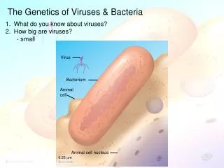
Virus. Bacterium. Animal cell. Animal cell nucleus. 0.25 m. The Genetics of Viruses & Bacteria. What do you know about viruses? How big are viruses? - small. The Genetics of Viruses & Bacteria. Capsomere of capsid. Membranous envelope. RNA. Capsomere. DNA. Head. Capsid.

E N D
Virus Bacterium Animalcell Animal cell nucleus 0.25 m The Genetics of Viruses & Bacteria • What do you know about viruses? • How big are viruses? • - small
The Genetics of Viruses & Bacteria Capsomereof capsid Membranousenvelope RNA Capsomere DNA Head Capsid Tail sheath DNA RNA Tail fiber Glycoprotein Glycoprotein 80 225 nm 18 250 mm 80–200 nm (diameter) 70–90 nm (diameter) 50 nm 20 nm 50 nm 50 nm (d) Bacteriophage T4 (a) Tobacco mosaic virus (b) Adenoviruses (c) Influenza viruses • What do you know about viruses? • How big are viruses? • What are the components of a virus? • Nucleic acid genome • Protein capsid
The Genetics of Viruses & Bacteria • What do you know about viruses? • How big are viruses? • What are the components of a virus? • How do viruses identify appropriate cells to infect? • Viruses bind to specific receptors • May cross species or be tissue-specific • 5. What is the lytic cycle of a bacteriophage?
Attachment. The T4 phage usesits tail fibers to bind to specificreceptor sites on the outer surface of an E. coli cell. Entry of phage DNA and degradation of host DNA.The sheath of the tail contracts,injecting the phage DNA intothe cell and leaving an emptycapsid outside. The cell’sDNA is hydrolyzed. 1 2 4 3 5 Release. The phage directs productionof an enzyme that damages the bacterialcell wall, allowing fluid to enter. The cellswells and finally bursts, releasing 100 to 200 phage particles. Phage assembly Synthesis of viral genomes and proteins. The phage DNAdirects production of phageproteins and copies of the phagegenome by host enzymes, usingcomponents within the cell. Assembly. Three separate sets of proteinsself-assemble to form phage heads, tails,and tail fibers. The phage genome ispackaged inside the capsid as the head forms. Head Tail fibers Tails Figure 18.6 The lytic cycle of phage T4, a virulent phage
The Genetics of Viruses & Bacteria • What do you know about viruses? • How big are viruses? • What are the components of a virus? • How do viruses identify appropriate cells to infect? • What is the lytic cycle of a bacteriophage? • What is the lysogenic cycle of a bacteriophage?
Phage DNA The phage attaches to a host cell and injects its DNA. Many cell divisions produce a large population of bacteria infected with the prophage. Phage DNA circularizes Phage Occasionally, a prophage exits the bacterial chromosome, initiating a lytic cycle. Bacterial chromosome Lytic cycle Lysogenic cycle Certain factors determine whether The bacterium reproduces normally, copying the prophage and transmitting it to daughter cells. The cell lyses, releasing phages. Prophage Lytic cycle is induced Lysogenic cycle is entered or New phage DNA and proteins are synthesized and assembled into phages. Phage DNA integrates into the bacterial chromosome,becoming a prophage. Figure 18.7 The lytic and lysogenic cycles of phage , a temperate phage
The Genetics of Viruses & Bacteria Glycoprotein Viral envelope Capsid RNA(two identicalstrands) Reversetranscriptase • What do you know about viruses? • How big are viruses? • What are the components of a virus? • How do viruses identify appropriate cells to infect? • What is the lytic cycle of a bacteriophage? • What is the lysogenic cycle of a bacteriophage? • How do retroviruses (like HIV) reproduce? • Reverse transcriptase – RNA back to DNA • Helper T cells
The virus fuses with the cell’s plasma membrane. The capsid proteins are removed, releasing the viral proteins and RNA. 1 HIV Membrane of white blood cell 2 Reverse transcriptase catalyzes the synthesis of a DNA strand complementary to the viral RNA. HOST CELL 3 Reverse transcriptase catalyzes the synthesis ofa second DNA strand complementary to the first. Reverse transcriptase Viral RNA RNA-DNAhybrid 4 The double-stranded DNA is incorporated as a provirus into the cell’s DNA. 0.25 µm HIV entering a cell DNA NUCLEUS Provirus ChromosomalDNA RNA genomefor the nextviral generation 5 Proviral genes are transcribed into RNA molecules, which serve as genomes for the next viral generation and as mRNAs for translation into viral proteins. mRNA 6 The viral proteins include capsid proteins and reverse transcriptase (made in the cytosol) and envelope glycoproteins (made in the ER). 7 Capsids are assembled around viral genomes and reverse transcriptase molecules. 8 Vesicles transport the glycoproteins from the ER to the cell’s plasma membrane. 9 New viruses bud off from the host cell. New HIV leaving a cell Figure 18.10 The reproductive cycle of HIV, a retrovirus
The Genetics of Viruses & Bacteria • What do you know about viruses? • How big are viruses? • What are the components of a virus? • How do viruses identify appropriate cells to infect? • What is the lytic cycle of a bacteriophage? • What is the lysogenic cycle of a bacteriophage? • How do retroviruses (like HIV) reproduce? • How do “new” viruses emerge? • Mutation of an existing virus since there is no proofreading • Spread of an existing virus from 1 host species to another • Spread of viral disease from a small isolated population • 9. What is the difference between horizontal & vertical transmission? • Horizontal – 1 organism spreads to another • Vertical – 1 organism inherits disease from parent • 10. What are viroids & prions? • Viroids – tiny molecules of naked, circular RNA that infect plants, • several hundred nucleotides long • Prions – infectious proteins (NO genetic material) • Slow incubation period – at least 10 yrs • Virtually indestructible • 1997 Nobel Prize in Medicine – Stanley Prusiner
Originalprion Prion Many prions Normalprotein Newprion Figure 18.13 Model for how prions propagate Mad cow disease Creutzfeldt-Jakob disease
The Genetics of Viruses & Bacteria • What do you know about viruses? • How big are viruses? • What are the components of a virus? • How do viruses identify appropriate cells to infect? • What is the lytic cycle of a bacteriophage? • What is the lysogenic cycle of a bacteriophage? • How do retroviruses (like HIV) reproduce? • How do “new” viruses emerge? • 9. What is the difference between horizontal & vertical transmission? • 10. What are viroids & prions? • 11. How is bacterial DNA different from eukaryotic DNA? • Bacterial Eukaryotic • Circular chromosome Linear chromosomes • Nucleoid region Nucleus • No introns (all exons) Introns & exons • Transcription coupled w/ translation Transcription & translation separate • How does bacterial DNA replicate its circular chromosome? • - Figure 16.16
Overall direction of replication Lagging strand Leading strand Origin of replication Helicase unwinds the parental double helix. 1 2 Molecules of single- strand binding protein stabilize the unwound template strands. The leading strand is synthesized continuously in the 5 3 direction by DNA pol III. 3 Leading strand Lagging strand OVERVIEW DNA pol III Leading strand 5 Replication fork DNA ligase DNA pol I 3 Primase 2 Parental DNA DNA pol III Lagging strand 1 Primer 3 4 Primase begins synthesis of RNA primer for fifth Okazaki fragment. 3 5 4 5 6 7 DNA pol III is completing synthesis of the fourth fragment, when it reaches the RNA primer on the third fragment, it will dissociate, move to the replication fork, and add DNA nucleotides to the 3 endof the fifth fragment primer. DNA ligase bonds the 3 end of the second fragment to the 5 end of the first fragment. DNA pol I removes the primer from the 5 end of the second fragment, replacing it with DNA nucleotides that it adds one by one to the 3’ end of the third fragment. The replacement of the last RNA nucleotide with DNA leaves the sugar- phosphate backbone with a free 3 end. Figure 16.16 A summary of bacterial DNA replication
The Genetics of Viruses & Bacteria • What do you know about viruses? • How big are viruses? • What are the components of a virus? • How do viruses identify appropriate cells to infect? • What is the lytic cycle of a bacteriophage? • What is the lysogenic cycle of a bacteriophage? • How do retroviruses (like HIV) reproduce? • How do “new” viruses emerge? • 9. What is the difference between horizontal & vertical transmission? • 10. What are viroids & prions? • 11. How is bacterial DNA different from eukaryotic DNA? • Bacterial Eukaryotic • Circular chromosome Linear chromosomes • Nucleoid region Nucleus • No introns (all exons) Introns & exons • Transcription coupled w/ translation Transcription & translation separate • How does bacterial DNA replicate its circular chromosome? • Figure 16.16 • Problem with circular chromosome?????? • Solved – topoisomerase
Table 16.1 Bacterial DNA replication proteins and their functions
Replicationfork Origin of replication Termination of replication Figure 18.14 Replication of a bacterial chromosome
The Genetics of Viruses & Bacteria • What do you know about viruses? • How big are viruses? • What are the components of a virus? • How do viruses identify appropriate cells to infect? • What is the lytic cycle of a bacteriophage? • What is the lysogenic cycle of a bacteriophage? • How do retroviruses (like HIV) reproduce? • How do “new” viruses emerge? • 9. What is the difference between horizontal & vertical transmission? • 10. What are viroids & prions? • 11. How is bacterial DNA different from eukaryotic DNA? • How does bacterial DNA replicate its circular chromosome? • Can bacterial cells do genetic recombination? • 3 (4) ways • Transformation – uptake of external DNA by a cell – Griffith • Transduction – phage transfers bacterial DNA • Conjugation – bacterial sex – direct transfer of genetic material • (Transposons)
Phage DNA B+ Phage infects bacterial cell that has alleles A+ and B+ A+ 1 Host DNA (brown) is fragmented, and phage DNA and proteins are made. This is the donor cell. A+ 2 B+ Donorcell A bacterial DNA fragment (in this case a fragment withthe A+ allele) may be packaged in a phage capsid. 3 A+ Crossingover Phage with the A+ allele from the donor cell infects a recipient A–B– cell, and crossing over (recombination) between donor DNA (brown) and recipient DNA (green) occurs at two places (dotted lines). 4 A+ A– B– Recipientcell The genotype of the resulting recombinant cell (A+B–) differs from the genotypes of both the donor (A+B+) and the recipient (A–B–). 5 A+ B– Recombinant cell Figure 18.16 Generalized transduction
1 m Sex pilus Figure 18.17 Bacterial conjugation
F Plasmid Bacterial chromosome F+ cell F+ cell Mating bridge F+ cell Bacterial chromosome F– cell A cell carrying an F plasmid(an F+ cell) can form amating bridge with an F– celland transfer its F plasmid. DNA replication occurs inboth donor and recipientcells, using the single parental strands of the F plasmid as templates to synthesize complementary strands. The plasmid in the recipient cell circularizes. Transfer and replication result in a compete F plasmid in each cell. Thus, both cells are now F+. A single strand of the F plasmid breaks at a specific point (tip of blue arrowhead) and begins tomove into the recipient cell. As transfer continues, the donor plasmid rotates(red arrow). 3 2 6 4 1 8 4 3 2 1 7 5 (a) Conjugation and transfer of an F plasmid from an F+ donor to an F– recipient Hfr cell F+ cell F factor The circular F plasmid in an F+ cellcan be integrated into the circularchromosome by a single crossoverevent (dotted line). The resulting cell is called an Hfr cell (for High frequency of recombination). B+ D+ C+ C+ A+ D+ A+ A+ B+ D+ D+ A+ C+ C+ B+ B+ A+ B+ C– C– C– C– F– cell B– B+ D– D– D– A+ B– B– D– B– A– A– A– A– A+ Since an Hfr cell has all the F-factor genes, it can form a mating bridge with an F– cell and transfer DNA. The location and orientation of the F factor in the donor chromosome determine the sequence of gene transfer during conjugation. In this example, the transfer sequence for four genes is A-B-C-D. A single strand of the F factorbreaks and begins to move through the bridge. DNA replication occurs in both donor and recipient cells, resulting in double-stranded DNA The mating bridgeusually breaks well before the entire chromosome and the rest of the F factor are transferred. (b) Conjugation and transfer of part of the bacterial chromosome from an Hfr donor to an F– recipient, resulting in recombination Temporary partial diploid C– C– Recombinant F– bacterium B+ B– D– D– B+ B– A– A– A+ A+ The piece of DNA ending up outside thebacterial chromosome will eventually be degraded by the cell’s enzymes. The recipient cell now contains a new combination of genes but no F factor; it is a recombinant F– cell. Two crossovers can result in the exchange of similar (homologous) genes between the transferred chromosome fragment (brown) and the recipient cell’s chromosome (green). Figure 18.18 Conjugation and recombination in E. coli Plasmid – extra-chromosomal, small, circular, self-replicating DNA
Figure 18.19 Transposable genetic elements in bacteria Insertion sequence 3 5 3 5 A T C C G G T… A C C G G A T… T A G G C C A … T G G C C T A … Transposase gene Inverted repeat Inverted repeat (a) Insertion sequences, the simplest transposable elements in bacteria, contain a single gene that encodes transposase, which catalyzes movement within the genome. The inverted repeats are backward, upside-down versions of each other; only a portion is shown. The inverted repeat sequence varies from one type of insertion sequence to another. Transposon Antibioticresistance gene Insertion sequence Insertion sequence 5 3 5 3 Transposase gene Inverted repeats (b) Transposons contain one or more genes in addition to the transposase gene. In the transposon shown here, a gene for resistance to an antibiotic is located between twin insertion sequences. The gene for antibiotic resistance is carried along as part of the transposon when the transposon is inserted at a new site in the genome.
Happy Thanksgiving!!!! • Have a safe & happy holiday break!! • Present pictographs when we come back • Test Tuesday • Review sessions Monday after school & Tuesday at 7
The Genetics of Viruses & Bacteria • What do you know about viruses? • How big are viruses? • What are the components of a virus? • How do viruses identify appropriate cells to infect? • What is the lytic cycle of a bacteriophage? • What is the lysogenic cycle of a bacteriophage? • How do retroviruses (like HIV) reproduce? • How do “new” viruses emerge? • 9. What is the difference between horizontal & vertical transmission? • 10. What are viroids & prions? • 11. How is bacterial DNA different from eukaryotic DNA? • How does bacterial DNA replicate its circular chromosome? • Can bacterial cells do genetic recombination? • How are metabolic pathways regulated? • Inhibition of enzyme activity – protein level • Inhibition of transcription – mRNA level
(b) Regulation of enzyme production (a) Regulation of enzyme activity Precursor Feedback inhibition Enzyme 1 Gene 1 Regulation of gene expression Gene 2 Enzyme 2 Gene 3 Enzyme 3 – Gene 4 Enzyme 4 – Gene 5 Enzyme 5 Tryptophan Figure 18.20 Regulation of a metabolic pathway
The Genetics of Viruses & Bacteria • What do you know about viruses? • How big are viruses? • What are the components of a virus? • How do viruses identify appropriate cells to infect? • What is the lytic cycle of a bacteriophage? • What is the lysogenic cycle of a bacteriophage? • How do retroviruses (like HIV) reproduce? • How do “new” viruses emerge? • 9. What is the difference between horizontal & vertical transmission? • 10. What are viroids & prions? • 11. How is bacterial DNA different from eukaryotic DNA? • How does bacterial DNA replicate its circular chromosome? • Can bacterial cells do genetic recombination? • How are metabolic pathways regulated? • Inhibition of enzyme activity – protein level • Inhibition of transcription – mRNA level • What is an operon? • A cluster of genes whose products function in a common pathway & • are regulated together • Repressible – usually on – tryptophan – trp operon - anabolic • Inducible – usually off – lactose – lac operon - catabolic
trp operon Promoter Promoter Genes of operon RNA polymerase Start codon Stop codon trpR trpD trpC trpB trpE trpA DNA Operator Regulatory gene 3 mRNA 5 mRNA 5 C E D B A Polypeptides that make up enzymes for tryptophan synthesis Inactiverepressor Protein (a) Tryptophan absent, repressor inactive, operon on. RNA polymerase attaches to the DNA at the promoter and transcribes the operon’s genes. Figure 18.21 The trp operon: regulated synthesis of repressible enzymes
DNA No RNA made mRNA Active repressor Protein Tryptophan (corepressor) (b) Tryptophan present, repressor active, operon off. As tryptophan accumulates, it inhibits its own production by activating the repressor protein.
Promoter Regulatorygene Operator DNA lacl lacZ NoRNAmade 3 RNApolymerase mRNA 5 Activerepressor Protein (a) Lactose absent, repressor active, operon off. The lac repressor is innately active, and inthe absence of lactose it switches off the operon by binding to the operator. Figure 18.22 The lac operon: regulated synthesis of inducible enzymes
lac operon DNA lacl lacz lacY lacA RNApolymerase 3 mRNA 5 mRNA 5' mRNA 5 -Galactosidase Permease Transacetylase Protein Inactiverepressor Allolactose(inducer) (b) Lactose present, repressor inactive, operon on. Allolactose, an isomer of lactose, derepresses the operon by inactivating the repressor. In this way, the enzymes for lactose utilization are induced.
Pictographs – a fun and creative representation of the process below Remember each group member must write a paragraph explaining how each symbol relates to the actual biological process. DNA replication Transcription Translation Mutations trp operon lac operon