Download
1 / 17
200 likes | 640 Vues
LECTURE 31- DIENCEPHALON AND PITUITARY GLAND . Dr. Mohammad Rehan Asad. At the end of the lecture the student should be able to. Identify the structure and divisions of diencephalon. Describe the boundaries of diencephalon. Discuss external and internal features of thalamus.

E N D
LECTURE 31- DIENCEPHALON AND PITUITARY GLAND Dr. Mohammad Rehan Asad
At the end of the lecture the student should be able to • Identify the structure and divisions of diencephalon. • Describe the boundaries of diencephalon. • Discuss external and internal features of thalamus. • Discuss sub thalamus, epithalamus, Habenular nucleus and pineal body. • Identify relations of hypothalamus. • Discuss hypothalamic nuclei in lateral and medial zones. • Identify afferent and efferent connections of hypothalamus. • Describe hypothalamic connections of pituitary gland.
DIENCEPHALON EPITHALAMUS THALAMUS SUBTHALAMUS HYPOTHALAMUS
Diencephalon • Extend from interventricular foramen to posterior commissure • Symmetrical right and left halves • Formed by hypothalamic structures and also include, from anterior to posterior, the optic chiasma, with the optic tract on either side; the infundibulum, with the tuber cinereum; and the mammillary bodies. • superior wall of the diencephalon is formed by the roof of the third ventricle • The medial surface: formed in its superior part by the medial surface of the thalamus and in its inferior part by the hypothalamus • lateral surface of the diencephalon is bounded by the internal capsule
Thalamus • large ovoid mass of gray matter that forms the major part of the diencephalon. • Present on each side of the third ventricle. • The anterior end of the thalamus is narrow and rounded and forms the posterior boundary of the interventricular foramen. • The posterior end is expanded to form the pulvinar, which overhangs the superior colliculus and the superior brachium. • The lateral geniculate body forms a small elevation on the under aspect of the lateral portion of the pulvinar.
Thalamus • The superior surface is covered medially by the telachoroidea and the fornix • laterally, it is covered by ependyma and forms part of the floor of the lateral ventricle • The inferior surface is continuous with the tegmentum of the midbrain • The medial surface forms the superior part of the lateral wall of the third ventricle and is connected to the opposite thalamus by a band of gray matter, interthalamic adhesion. • The lateral surface is separated from the lentiform nucleus internal capsule
Epithalamus • consists of the habenular nuclei and their connections and the pineal gland • Habenularnucleus is a small group of neurons situated just medial to the posterior surface of the thalamus. • It is a centre for integration of olfactory, visceral, and somatic afferent pathway • The pineal gland is a small, conical structure that is attached by the pineal stalk to the diencephalon. • The base of the pineal stalk possesses a recess that is continuous with the cavity of the third ventricle • The superior part of the base of the stalk contains the habenularcommissure • The inferior part of the base of the stalk contains the posterior commissure.
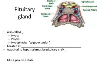
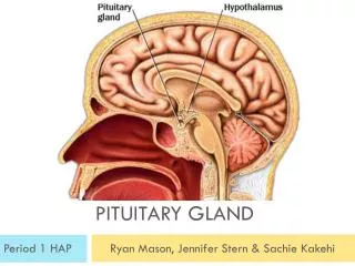
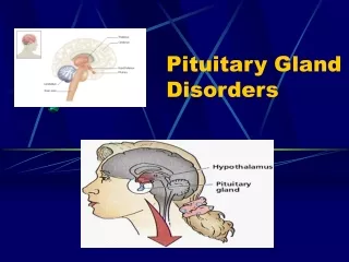
Hypothalamus • Extends from the region of the optic chiasma to the caudal border of the mammillary bodies • Located below the hypothalamic sulcus on the lateral wall of the third ventricle. • The hypothalamus controls and integrates the functions of the autonomic nervous system and the endocrine systems and plays a vital role in maintaining body homeostasis. • It is involved in such activities as regulation of body temperature, body fluids, drives to eat and drink, sexual behaviour, and emotion.
Relations • Anterior: area that extends forward from the optic chiasma to the lamina terminalis and the anterior commissure • Referred as the preoptic area. • Caudally, the hypothalamus merges into the tegmentum of the midbrain. • The thalamus lies superior to the hypothalamus • the subthalamic region lies inferolaterally to the hypothalamus
Nuclei • Medial Zone • part of the preoptic nucleus; the anterior nucleus, which merges with the preoptic nucleus; part of the suprachiasmatic nucleus; the paraventricular nucleus; the dorsomedial nucleus; the ventromedial nucleus; the infundibular (arcuate) nucleus; and the posterior nucleus.
Nuclei • Lateral Zone • part of the preoptic nucleus; part of the suprachiasmatic nucleus ; the supraoptic nucleus; the lateral nucleus ; the tuberomammillary nucleus; the lateral tuberal nuclei.
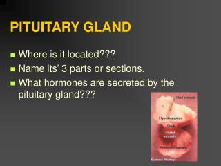
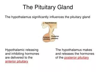
Connections of the Hypothalamus With the Hypophysis Cerebri Connected to the hypophysiscerebri (pituitary gland) by two pathways: (1) nerve fibers that travel from the supraoptic and paraventricular nuclei to the posterior lobe of the hypophysis and
Connections of the Hypothalamus With the Hypophysis Cerebri • Long and short portal blood vessels that connect sinusoids in the median eminence and infundibulum with capillary plexuses in the anterior lobe of the hypophysis. • These pathways enable the hypothalamus to influence the activities of the endocrine glands.