Download
1 / 24
260 likes | 418 Vues
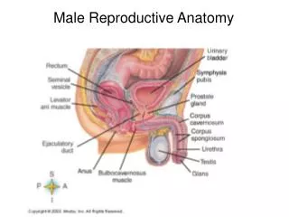
Male and Female Reproductive Anatomy. Healthy Active Living. Male Reproductive System. Testes.

E N D
Male and Female Reproductive Anatomy Healthy Active Living
Testes • The paired oval testes, also known as the male gonads, hang in the scrotal sac. Usually the right testis hangs higher than the left one by about 1 cm. The testes hang outside the body because the temperature inside the body is too high to produce sperm, so they are produced in the testes at about 3 °C lower than body temperature.
Epididymis • At the back of the olive-shaped body of each testis is a cap formed by the many coils of a 20-foot long tube called the epididymis. The function of the epididymis is to collect the immature sperm from the testis. • As the sperm make their long journey through the epididymis they become mature sperm. This journey takes about 20 days and during its course the sperm become fertile and they also become able to move in a swimming motion (doctors refer to the sperm then as ‘motile’).
Vas deferens • Joined to the epididymis is the vas deferens — a thick walled tube which transports sperm from the epididymis up to the prostate gland. The section of the vas deferens that is above the testis can be felt through the loose part of the scrotum. When a vasectomy is performed, it is this part of the vas deferens that is snipped. • The vas deferens empties into the ejaculatory duct, which passes through the prostate gland to merge with the urethra.
The urethra • The urethra serves as the tube down which urine passes from the bladder through the penis to the outside and also the tube down which semen is ejaculated.
The prostate • The prostate is a walnut-shaped gland that surrounds the urethra. Along with the seminal vesicles it produces the fluid secretions that support and nourish the sperm. Without this fluid to dilute them the sperm cannot move easily. • After the age of 40 the prostate enlarges and can press on the urethra. An enlarged prostate is often the cause of urinary problems in older men.
Glans • The sensitive bulbous structure at the distal end of the penis • Typically, the glans is completely or partially covered by the foreskin except by men who have been circumcised.
Bladder • The urinary bladder is a muscular sac in the pelvis, just above and behind the pubic bone. When empty, the bladder is about the size and shape of a pear. • Urine is made in the kidneys, and travels down two tubes called ureters to the bladder. The bladder stores urine, allowing urination to be infrequent and voluntary. The bladder is lined by layers of muscle tissue that stretch to accommodate urine. The normal capacity of the bladder is 400 to 600 mL. • During urination, the bladder muscles contract, and two sphincters (valves) open to allow urine to flow out. Urine exits the bladder into the urethra, which carries urine out of the body. Because it passes through the penis, the urethra is longer in men (8 inches) than in women (1.5 inches).
Scrotum • The scrotum is the protective sac of skin and muscle that contains the testicles, the epididymis and part of the spermatic cord. • The epididymis is where the sperm created in the testes is stored. The spermatic cord is made up of blood vessels, nerves, lymphatic vessels and the vas deferens. The spermatic cord has layers of tough tissue to protect it from damage. The vas deferens is the tube that carries the sperm between the testes and epididymis to the urethra
Rectum • The rectum is about eight inches long and serves, basically, as a warehouse for poop. It hooks up with the sigmoid colon to the north and with the anal canal to the south • The rectum has little shelves in it called transverse folds. These folds help keep stool in place until you're ready to go to the bathroom. When you're ready, stool enters the lower rectum, moves into the anal canal, and then passes through the anus on its way out.
Urethral Opening • The male urethra is a narrow fibromuscular tube that conducts urine and semen from the bladder and ejaculatory ducts, respectively, to the exterior of the body. Although the male urethra is a single structure, it is composed of a heterogeneous series of segments: prostatic, membranous, and spongy.
Foreskin • The fold of skin which covers the head (the glans) of the penis. Also called the prepuce. • The foreskin is thus not fully separable from the glans in about 96% of newborn boys. By a year of 1 year, the foreskin can be retracted in 50% of boys and by 3 years, the foreskin can be retracted in 80% to 90% of uncircumcised boys.
Seminal Vesicles • The seminal vesicles are paired organs of the male genitourinary system. Specifically, they are genital organs. The male genital organs include the penis, testes, excretory genital ducts, ductus deferens, seminal vesicles, prostate, and bulbourethral glands. Of these organs, the seminal vesicles, prostate, and bulbourethral glands are considered accessory glandular structures. • These organs work together to produce and excrete semen
Anus • The anus is the opening where the gastrointestinal tract ends and exits the body. The anus starts at the bottom of the rectum, the last portion of the colon (large intestine). The anorectal line separates the anus from the rectum. • Tough tissue called fascia surrounds the anus and attaches it to nearby structures. • Circular muscles called the external sphincter ani form the wall of the anus and hold it closed. Glands release fluid into the anus to keep its surface moist. • A plate-like band of muscles, called the levatorani muscles, surround the anus and form the floor of the pelvis. A network of veins lines the skin of the anus.
Inner Labia • two thin folds of skin between the labia majora, extending from the clitoris backward on both sides of the vaginal orifice, ending between it and the labia majora. Anteriorly each labium divides into an upper and a lower division. The upper divisions pass above the clitoris and meet to form the preputiumclitoridis. The lower divisions pass beneath the clitoris and unite to form the frenulum of the clitoris
Hymen • A hymen is the thin piece of tissue that partially blocks the entrance to the vagina. It is sometimes called the maidenhead or cherry • Although some women are born without a hymen, most have one, and the hymen varies in size and shape from woman to woman. The hymen usually does not cover the entire vaginal opening, since there must be some way for the menstrual fluid, or period, to leave the body
Vagina • The vagina is an elastic, muscular canal with a soft, flexible lining that provides lubrication and sensation. The vagina connects the uterus to the outside world. The vulva and labia form the entrance, and the cervix of the uterus protrudes into the vagina, forming the interior end. • The vagina receives the penis during sexual intercourse and also serves as a conduit for menstrual flow from the uterus. During childbirth, the baby passes through the vagina (birth canal).
Cervix • The cervix is a cylinder-shaped neck of tissue that connects the vagina and uterus. The cervix is made of cartilage covered by smooth, moist tissue, and is about 1 inch across. There are two main portions of the cervix: • The part of the cervix that can be seen from inside the vagina during a gynecologic examination is known as the ectocervix. An opening in the center of the ectocervix, known as the external os, opens to allow passage between the uterus and vagina. • The endocervix, or endocervical canal, is a tunnel through the cervix, from the external os into the uterus. • The cervix produces cervical mucus that changes in consistency during the menstrual cycle to prevent or promote pregnancy.
Uterus • A hollow, pear-shaped organ that is located in a woman's lower abdomen, between the bladder and the rectum. The narrow lower portion of the uterus is the cervix (the neck of the uterus). • the inner layer (endometrium) of the uterus goes through a series of monthly changes known as the menstrual cycle. Each month, endometrial tissue grows and thickens in preparation to receive a fertilized egg. (ovulation)
Ovaries • The female gonad, one of a pair of reproductive glands in women. The ovaries are located in the pelvis, one on each side of the uterus. Each ovary is about the size and shape of an almond • The ovaries produce eggs (ova) and female hormones. During each monthly menstrual cycle, an egg is released from one ovary. The egg travels from the ovary through a fallopian tube to the uterus. The ovaries are the main source of female hormones, which control the development of female body characteristics, such as the breasts, body shape, and body hair. They also help regulate the menstral cycle and pregnancy.
Fallopian Tube • The uterine tube (Fallopian tube) carries an egg from the ovary to the uterus. • Women who no longer want children can have their "tubes tied" to prevent eggs from moving from the Fallopian tubes into the uterus
Sources: • http://www.medterms.com/script/main/art.asp?articlekey=4705 • http://www.globalrph.com/medterm.htm • http://www.getbodysmart.com/ap/site/resourcelinks/links.html