Download
1 / 13
130 likes | 247 Vues
This study focuses on the hierarchical levels and composition of bone, particularly the lamellar structure, using various microscopy techniques. Investigating bone lamellae helps understand its formation and predict macroscopic behaviors. The research aims to provide insights into bone's mechanical properties and internal organization.

E N D
Lamellar Characterization of Bone Presented by: Melanie Patel Advisors: Dr. Surya Kalidindi Dr. Haviva Goldman
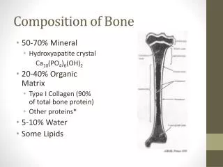
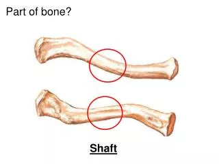
Definition • Bone is a stiff skeletal material made principally of the fibrous protein collagen, impregnated with a mineral closely resembling calcium phosphate. • A composite material consisting of fibrous protein, collagen, stiffened by extremely dense filling and surrounding of calcium phosphate crystals. • The mineralized collagen fibrils are considered the basic building block of the bone.
Collagen • Basic building block of collagenous framework is a triple helical molecule • 3 amino acids: glycine, proline, hydroxyproline. • The structure follows the sequence of Glycine-X-Y where X is proline and Y can be either proline or hydroxyproline. • Hodge and Petruska Model
Purpose • Characterize Lamellar Structure • Quantitative • Orientation • Chemical Make-up • Size (Thickness) • Use information to predict Macroscale Properties.
Lamellae • The individual lamellae of bone consist of a fibril matrix with collagen fibers interweaving each other • Thin Lamellae • dense, fiber-rich, lack substance • 1-3 μm • Thick Lamellae • Collagen-poor layer, more substance • 2-4 μm
Theories • Plywood Structure- orthogonally alternating layers • Fiber orientation at the Boundary- Ascenzi and Benvenuti, fiber directions do not change abruptly • 3-D Organization- Marotti • Twisted Plywood- gives rise to arc pattern in osteons • Rotated Plywood
Microscopy • Polarized Light • SEM • TEM
Characterization • Nanoindentation • AFM • EDAX??
Experimentation thus far • Femoral bone sample obtained • Samples mounted in PMMA solidified • Cut, grind, polish • Light Microscopy • Nanoindentation, AFM • EDAX Experimentation plans
Sources • Curry, John D. “Bones: Structure and Mechanics”. 2002. • Weiner S. and Wagner H.D. “The Material Bone: Structure-Mechanical Function Relations”. Annu. Rev. Mater. Sci. (28). 1998: 271-298. • Weiner, Stephen and Traub, Wolfie. “Bone Structure: from angstroms to microns” Faseb Journal. (6) 1992: 879-885. • Weiner S. et al. “Rotated Plywood Structure of Primary Lamellar Bone in the Rat: Orientations of the Collagen Fibril Arrays” Bone. (20) 1997: 509-514. • Marotti, Gastone and Muglia, Maria Antonietta. “A scanning electron microscope study of human bone lamellae. Proposal for a new model of collagen lamellar organization.” Arch. Ital. Anat. Embroil. (93) 1988: 163-175. • Giraud-Guille, M.M. “Twisted Plywood Architecture of Collagen Fibrils in Human Compact Bone Osteons” Calcif Tissue Int. (42) 1988: 167-180. • Ziv, V. et al. “Microstructure-Microhardness Relations in Parellel-Fibered and Lamellar Bone” Bone. (18) 1996: 417-428. • Ascenzi A. and Benvenuti A. “Orientation of Collagen Fibers at the Boundary Between Two Successive Osteonic Lamellae and its Mechanical Interpretation” J. Biomechanics. (19) 1986: 455-463. • Bromage, Timothy G. et al. “Circularly Polarized Light Standard for Investigations of Collagen Fiber Orientation in Bone” The Anatomical Record. (274B) 2003: 157-168. • Rubin, Matthew et al. “TEM analysis of the nanostructure of normal and osteoporotic human trabecular bone” Bone. (33) 2003: 270-282.