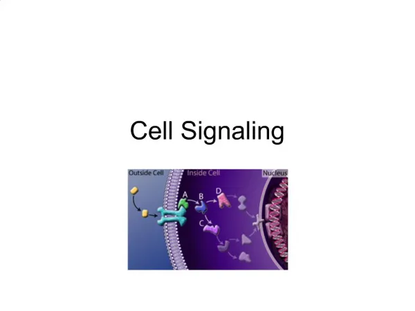
Cell Signaling (BIO-203)
Cell Signaling (BIO-203). Lecture 3. Types of G proteins. Humans have 21 different G α subunits 6 G β subunits 12 G γ subunits Different G βγ function similarly. GPCR that regulate ion channels.

Cell Signaling (BIO-203)
E N D
Presentation Transcript
Cell Signaling (BIO-203) Lecture 3
Types of G proteins • Humans have 21 different Gα subunits • 6 Gβ subunits • 12 Gγ subunits • Different Gβγ function similarly
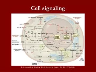
GPCR that regulate ion channels • The simplest cellular responses to a signal is the opening or closing of ion channels essential for transmission of nerve impulses • Nerve impulses are essential to the sensory perception of environmental stimuli (light, odor) to transmission of information to and from the brain and to the stimulation of muscle movement • During transmission of nerve impulses, the rapid opening and closing of ion channels causes changes in the membrane potential • Some neurotransmitter receptors are GPCRs whose effector proteins are Na or K channels
Neurotramsmitter binding to these receptors causes the associated ion channel to open or close leading to changes in the membrane potential e.g acetyl choline involved in K transport
http://highered.mcgraw-hill.com/sites/0072943696/student_view0/chapter10/animation__membrane-bound_receptors__g_proteins__and_ca2__channels.htmlhttp://highered.mcgraw-hill.com/sites/0072943696/student_view0/chapter10/animation__membrane-bound_receptors__g_proteins__and_ca2__channels.html
The alpha subunit of the G-protein is activated by.. A)separating from the gamma and beta subunits. B)the G-protein changing conformation. C)binding to the calcium ions D)replacing the GDP with GTP. E)replacing the GTP with GDP. • The activated alpha subunit then binds to... A)the calcium ion channel B)the calcium ions. C)the gamma and beta subunits. D)GDP E)GTP • As a result... A)the calcium channel opens and calcium ions leave the cell B)the calcium channel opens and calcium ions enter the cell. C)the calcium channel closes. D)the calcium ions bind to calmodulin. E)the calcium ions bind to the ligand receptor site. • The G-protein changes conformation when the GTP replaces the GDP on the alpha subunit. A)True B)False • The combination of the calcium and the calmodulin produces the response of the cell to the ligand. A)True B)False
Answers • D • A • B • B • A
Acetylcholine receptors in the heart muscle activate a G Protein that opens K+ Chanel • Activation of acetylcholine receptors in cardiac muscle slows the rate of heart muscle contraction. • It is coupled to a complex of Gαi-Gβγ protein. • Binding of acetylcholine triggers activation of Gαi subunit followed by its dissociation from the Gβγ subunit. • The released Gβγ subunit binds to and opens a K+ channel. • The increase in K+ permeability hyperpolarizes the membrane which reduces the frequency of heart muscle contraction.
Acetylcholine receptors in the heart muscle activate a G Protein that opens K+ Chanel
Hyperpolarization • It is a change in a cell's membrane potential that makes it more negative. It is the opposite of a depolarization. • It is often caused by efflux of K+through K+ channels, or influx of Cl– through Cl– channels. On the other hand, influx of cations, e.g. Na+ through Na+ channels or Ca2+ through Ca2+ channels inhibits hyperpolarization. • The efflux of K+ ions from the cytosol increases inside-negative ion potential across the plasma membrane that lasts for several seconds. it reduces the frequency of muscle contraction.
Light activates Gαt- Coupled rhodopsins • Human retina contains 2 types of photoreceptor cells: • Rods stimulated by moonlight over a range of wavelengths. • Cones involved in color vision. • They are the primary recipients of visual stimulation. • Rhodopsin consists of the protein opsin which has a usual GPCR structure covalently bonded to light-absorbing pigment 11-cis-retinal.
The trimeric G Protein couple to rhodopsin is called transducin (Gt). • It contains Gαt subunit. • Rhodopsin and Gαt subunit are found only in rod cells.
Rhodopsin is sensitive enough to respond to a single photon of light, this response takes place in the form of isomeriztion, • Therefore, when a photon of light enters the eye, it is absorbed by the retinal and causes a change in its configuration • from 11-cis retinal to all-trans retinal. • This isomeriztion induces conformational changes in Rhodopsin that activates the G-protein.
Light activated rhodopsin pathway • In dark adopted rod cells: • Light absorption generated activated opsin • Opsin binds inactive GDP-bound Gαt protein and mediates replacement of GDP with GTP • The free Gαt-GTP activates cGMPphosphodiesterase (PDE) by binding to its inhibitory γ subunits and dissociating them from the catalytic α and β subunits. • The free α and β subunits convert cGMP to GMP. • The resulting decrease in cGMP leads to dissociation of cGMP from the nucleotide-gated channels in the plasma membrane and closing of channels. • The membrane then becomes hyperpolarized.