Download
1 / 47
470 likes | 552 Vues
Explore the complexities of human genetics, inherited disorders, and the various inheritance patterns in monogenic, polygenic, and multifactorial diseases. Learn about the Human Genome Project's impact on genetic research.

E N D
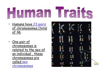
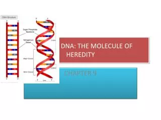
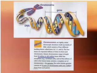
Each of the 46 chromosomes of humans is made up of single molecule of double-stranded DNA • if stretched out, the DNA from a single cell would extend approximately 2 meters in lenght • an elaborate system of coling, which also seem to be involved in the control of gene expression is present in memmalian cells • basic proteins called histones provide a core around which DNA is wound in a double loop composing approximately 146bp of DNA - „NUCLEOSOME“ • the resulting „beads-on-string“ DNA structure results in a compaction of lenght of about a factor of 7.Further organisation occurs by arrangement of the nucleosomes in solenoid fashion and by higher order structural complexities.
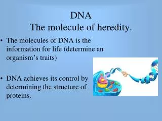
The human genome- the total genetic information (DNA content) in human cells. • It comprise two genomes: • a nuclear genome • mitochondrial genome
The human nuclear and mitochondrial genomes • differ in many aspects of their organisation and expression
Inherited disorders It is thought that about 5% of all newborn babies come into the world with an inherited disorder. About 0.5% have a clinically relevant chromosome anomaly and about 1% have a monogenic inherited disorder. The remainder of disorders are multifactorialor due to external factors. Among those who die before their 65th year, inherited disorders are still the fifth most frequent cause of death. Most deaths result from inherited heart disorders followed by anomalies of the central nervous system (brain,spinal cord) as well as urogenital anomalies (urinary and reproductive organs) and gastrointestinal anomalies (digestive organs).
In monogenic diseases, only a single gene is altered (mutant) with the consequence that the pattern for a specific protein is flawed, which in turn leads to the manifestation (development) of a disease. Sickle cell disease is an example of this. Monogenic diseases are often rare and severe illnesses. At present, over 6,000 genes are known whose mutations lead to various monogenic disorders. Currently, a molecular genetics analysis can be made on 1,000 of these diseases.
With polygenic diseasesit is the interaction of several gene alterations (mutations) which leads to the development of an illness. • the mutation of a single gene has a much less serious effect than with a monogenic disease. • another important difference to monogenic diseases is that polygenic diseases are very common in the population. Apart from the mutation of several genes, one or more environmental factors usually contribute to the manifestation of these diseases (multifactorial diseases). Onset, severity and course of these illnesses are also significantly influenced by environmental factors (e.g. nutrition, exercise). • For this group of illnesses, the contribution of the gene can be thought of as a “predisposition”. At present, there are only very few illnesses for which the contributory gene has been identified: e.g. in the case of thrombotic diseases (APC [Activated Protein C] resistance).
Genes and diseases The boundaries between monogenic, polygenic and multifactorial diseases can be expressed by the following statement: genes can always be represented as more or less strongly predisposing factors for the development of an illness.
Inheritance rules for genetic diseases • The following inheritance processes are known for monogenic diseases. In order to understand these rules of inheritance it is important to emphasize that : • every genein the cell nucleusis expressed twice– in the same way as all chromosomes are expressed twice (one set from the maternal and one set from the paternal parental set). • What is decisive for the onset of a genetic disease is whether a mutated genecan prevailin its pathological action against its “healthy” counterpart(gene homologue) or whether the other gene also has to be mutated. These inheritance patterns were already observed in the 19 century byGregor Mendelin his famous cross-breeding experiments with peas, which form the basis for today's comprehensive family tree analyses.
Autosomal dominant inheritance process In this inheritance pattern only one of the two homologous genes is mutated and although another normal gene is present (heterozygosity), the illness still appears (dominant gene effect). If, therefore, one of the parents carries this gene, there is a 50% probability that it will be transmitted to each child. Both men and women can be affected by this. This inheritance pattern accounts for over 60% of monogenic diseases,representing by far the most common inheritance process. This inheritance pattern is followed by many diseases which exhibit changes in the proteins forming the basis for connective and supporting tissue (e.g. Marfan’s syndrome, achondroplasia, hypertrophic cardiomyopathy). Obviously a mutated protein in just half the amount will have a pathological effect on the human organism in such cases.
Autosomal recessive inheritance • In this inheritance pattern, both homologous genes must be mutated (homozygosity) in order to produce an illness in the affected person. • The individual must therefore have inherited a corresponding gene mutation from both the father and the mother. • Individuals, who only receive one version of the mutated gene are called carriers. • Both sexes can be affected. If, for example, both parents are carriers, there is a 25% chance that the child will receive both mutated genes and so develop the • illness. Many metabolic diseases fall into this category (e.g. cystic fibrosis, phenylketonuria, adrenogenital syndrome, haemochromatosis). In the heterozygote state (i.e. only one gene copy is mutant) it seems that the human organism can often compensate for this state and make do with half of the unchanged protein. Only when both gene copies are mutated does the amount of faulty enzymes have a pathological effect which can then no longer be compensated.
X chromosome inheritance (sex-linked inheritance) Women have two X chromosomes. If they have a recessively acting mutated gene on one X chromosome, they are carriers for the corresponding illness. Men have only one X chromosome, since the other sex chromosome is a Y chromosome. If they have the mutated gene on the X chromosome, they will develop the illness as a rule. If a woman is a carrier for the illness inherited by the X chromosome, there is a 50% chance that she will pass on this illness to her son. Her daughters have a 50% chance of becoming a carrier for this illness.
Inheritance process in polygenic diseases Of course, in these cases too the individual genes take part in the above mentioned inheritance processes. However, there are always several genes involved in causing polygenic diseases with different types of interaction possible. Sometimes the effect of several genes must be added together and at other times a certain threshold has first to be crossed before an illness becomes manifest. Often individual genes have a complex relationship with each other (control and regulation genes in a gene network). In addition, one or more environmental factors may contribute to the manifestation of the corresponding illness. Given the large number of causative factors and their interrelationships, it is easy to understand thatthere are also a large number of variations in the genesis of multifactorial diseases.These are expressed in different ages of onset of the illness, different courses of the illness and different degrees of severity. In general, there is a fluid transition from the healthy state to the pathological state in polygenic diseases if there is no threshold effect. Thisbreadth in the variation of mutations leading to the same illness makes it impossible to make a prediction of symptoms and prognosis with current scientific knowledge.
Identification of inherited diseases • ) Phenotype analysis • The simplest and for centuries the most common type of genetic analysis • is the analysis of the so-called phenotype. The analysis involves the end • product of the genes which are translated into proteins by transcription • and translation.These proteins determine the way an individual’s body • looks. • Thefollowing points apply to phenotype analyses (here specifically: biochemical investigations): • Genes are directly responsible for the production of hormones, enzymes and other proteins. To this extent, the analysis of these substances and metabolic products can also be classified as a genetic • test in a wider sense. • Indication: Especially in the case of metabolic diseases in newborn babies. • Investigation procedure: Diagnostic measurement of altered or missing proteins using blood or urine analysis. This provides indirect evidence of a mutation of the gene responsible for this. • Examples: Phenylketonuria, alpha1-antitrypsin deficiency • There is clinical evidence of a connection between severe alpha1-antitrypsin deficiency and liver cirrhosis as well as lung emphysema. There are several different courses of the illness. In the early childhood form, in which liver cirrhosis and lung emphysema occur in infancy, the children usually die from the complications before their twelfth year. Another form primarily affects adults, in which emphysema with a rapidly progressive course occurs before their 40th year. Environmental • factors (nicotine, dust) play an important part in promoting the manifestation of this illness.
) Chromosome analysis (cytogenetic investigations) • This includes microscope examinations to investigate chromosome • alterations in terms of number (duplication or loss of individual chromosomes • = numeric chromosome aberration) and in terms of structure • (wrong composition, chromosome breaking = structural chromosome • aberration). There is no detailed investigation of individual genes in • such cases. • Indication: Anomalies in children (malformations, retarded development) • in the context of prenatal diagnosis, tendency to miscarriages, infertility. • A relatively new technique is molecular cytogenetics, which uses a combination of cytogenetic and molecular genetics methods. • Fluorescence-labelled DNA sequences are often used as diagnostic “probes”, hence the designation “FISH” (Fluorescence in situ hybridization)
C) Molecular genetics testing (DNA analysis, genome analysis DNA tests) • This provides evidence of a gene mutation responsible for producing • the illness. Here it is determined whether the sequence of the DNA bases • (nucleotide sequence) has changed within the affected • DNA/RNA diagnosis of genetic diseases • Not all mutation test use DNA . Testing RNA by RT-PCR has advantages when screening genes with many exons ( NF1 gene, DMD gene...) or seeking splicing mutations. • Very important in molecular genetic testing is using a protein-based functional assay, which may classify the products into two simple groups: functional and nonfunctional – essential question in most diagnostics
Limitations of DNA analysis • It has long been known from research into identical twins that monogenic and also polygenic diseases sometimes do not occur in both twins, even though the genetic information is the same in identical twins. So it is not surprising either that in monogenic diseases in which only a single gene is altered, the phenotypic consequences are often difficult to predict even in the case of a positive test result. This is due to several factors: • Penetrance: not every pathogenic mutation leads to the manifestation of a disease in the lifetime of a person. For example, the gene mutation for neurofibromatosis is manifested in almost all affected individuals. In inherited ovarian cancer this proportion is considerably reduced. This can be described as “reduced penetrance”. • Expressivity on the other hand describes quantitative differences in the manifestation of the disease/symptoms. Sometimes, the two concepts are difficult to separate, when, for example, a disease is so weakly manifested that it can no longer be diagnosed. The expressivity can fluctuate strongly especially in dominant monogenic diseases (e.g. Marfan’s syndrome, neurofibromatosis). • The age at which the disease manifests itself can vary strongly. An example of this is Huntington’s chorea. Differences in the onset of diseases are sometimes explained by so-called dynamic mutations. In passing on to the next generation, the disease-inducing mutation can lead to an earlier onset of the illness (anticipation) involving the extension of a mutated sequence of bases. • In many cases, genetic information is manifested in a different way when it is inherited from the mother than when it is inherited from the father. Here one speaks of imprinting.
Indirect DNA analysis gene CFTR - intron 8 - polymorphic site (CA)n GTATCACACACATTCGG from father from mother chr.7 allele A1: ------ GTATCACATTCGG---- the lenght of this allele is 130 bp allele A2: -----GTATCACACACATTCGG--- the lenght of this allele is 134 bp
chr.7 dF508 / non non / ? A1 / A3 A1 / A2 A1 / A3 A1 / A1 dF508 / ? non / non A1 / A2 A1 / A3 informative A1 / A1 A1 / A3 informative mutation in CFTRgene chr.7 chr.7 chr.7
dF508 / non non / non A1 / A3 A2 / A5 dF508 / non non / non dF508 / G542X A1 / A2 A3 / A5 A1 / A4