Download
1 / 65
660 likes | 970 Vues
HEART & BLOOD VESSELS ( Angiolog y ). by : Gracia Angelina Hendarti, MSi, Drh. HEART & BLOOD VESSELS (Angiolog y ). Is the description of the organs of circulation of the blood & lymph, the heart and vessels, including the spleen and thymus. c ardiovas c uler system :

E N D
HEART & BLOOD VESSELS (Angiology) by: Gracia Angelina Hendarti, MSi, Drh
HEART & BLOOD VESSELS (Angiology) • Is the description of the organs of circulation of the blood & lymph, the heart and vessels, including the spleen and thymus. • cardiovasculer system: heart( latin :COR) blood vessels: arteries & capillaries, veins • Lymphatic system : lymphatic organ lymphnodes lymph vessels
Pericardium • Is the fibroserous sacwhich encloses the heart, in part, great vessels connected with it. • fibrous layer (pericardium fibrosum): relative thin, but strong and inelastic. • pericardium serosum : is a closed sac, surrounded by the fibrous pericardium and invaginated by the heart. It is smooth and glistening and contain a small amount of clear serous fluid : liqour pericardii. • Pericardium visceralis/lamina visceralis/epicardium • Pericardium parietalis/lamina parietalis • Pericardium visceralis : covers the heart and parts of the great vessels. • Pericardium parietalis : lines the fibrous layer, to which it is closely attached.
Pericardium blood vessel Pericardium Visceralis (epicardium) Cavum pericardium heart Pericardium Parietalis Pericardium Fibrousa Sternopericardiaca ligament
PERICARDIUM • It is attached dorsally to the large vessels at the base of the heart and is continued in part up to the longus colli muscle. • Ligamentum sternopericardiaca : it is a strong ligament attached the pericardium ventrally to the middle of the caudal half of the thoracic surface of the sternum. ( in cattle, horse and swine) and in carnivore by a phrenopericardiac ligament to the diaphragm.
Wall / cardiac muscles : Epicardium Myocardium endocardium heart
Heart = cor Atrium dexter Auricula sinister Ventrikel dexter Ventrikel sinister
Heart = cor R L vessel blood vessel atrium auricula atrium valve tricuspidalis valve mitralis ventricle left ventricle Septum interventricularis
Valvula of the Heart Right atrioventricular valve (valvula tricuspidalis) left atrioventriculularis valve (valvula bicuspidalis/mitralis)
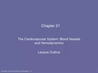
BLOOD VESSELS • Arteri : • Leave he heart, • recieve & sent the blood from the heartto the tissue • contain of O2 except in the arteria Pulmonalis • position : usually more deeper than vena(profunda) • Structure : more elasticthan vena • the pressure more stronger than venae • Vena : • the blood go to he heart. • Receive bloodfrom the tissue and send to the heart • contain metabolism product (especially CO2) except v.pulmonalis • location : more superficial • have valve • Structure : the wallis thinner and not elastic • the pressure is weaker
Structureof the blood vessels • The wall of the blood vessel compose of : • First layer : tunica interna/intima : • Endothel • Membrane elastica interna (subendothel) • Second layer : tunica media : • thicker part, containelastic tissue andsmooth muscle • Third layer: tunica adventitia : • Compose of fibrous and fibroareolar tissue
BLOOD VESSELS STRUCTURE endothel Tunica interna Membrane elastica interna Tunica media Tunica adventitia
Vein arrow indicates the direction of blood flow. valve Most veins are also distinguish by the presence of valves, which are repeated at interval along their length; the valves ensure a unidirectional flow and prevent reflux of blood when the circulation stagnates. Each valve consists of two or three semilunar cusps facing each other
BLOOD VESSEL COMPOSITION according to the thickness of the tunica media ( elastic tissue) : the smaller the diameter of the blood vessel, tunica media become thinner and thinner • Artery : • Aorta • Artery • Arteriol • capiler • Sinusoid • Vein : • vein • Venule
Artery : • Aorta : the biggest diameter , leave the cor, accept the sistolic output from ventricle • Artery : branch of the aorta • Arteriol: smaller diameter, a litter biggerthan capiler, functionto adjust blood stream and to pump the perifer blood. • Capiler : • Endothel is thin • Supporting tissue is thinner too, • Place for the blood enter to the interstitial tissue • Parts of the liquid which contain metabolic waste is absorbed by venule. • On special organ, endothelialcapiler which enterto the organ have pore. Ex : on the intestine villiand glomerulus
ARTERIES • the arteries may be distinguished from other vessels by their white, thick, and relatively rigid walls and their empty lumen (unless filled with an injection mass for the convenience of the dissector).
Sinusoid : parts of thecapiler which can be find in special organs. Ex: lever, spleen and medulla spinalis. specificity: usually inside the organs • Vein : • Vein : thinner wall, their frequently collapsed appearance, and their capacity, which is invariably greater than that of the associated arteries.in the large diameter of the vena the anatomic structure almost the same asthe arteri. • Very largest arteries and veins run separately, but most veins of medium and lesser size accompany the corresponding arteries to which they are said to be satellite • Venule : vena ending, smallest vena,tunica interna always thin and do not have membrane elastic interna
CAPILER closed capiler open capiler Arteriol venule spinchter
Anastomose : direct connection between arteri and vein without passing thecapiler in order toshorten the connection between arteri and vein, • Fx : • Passing the blood stream from the arteri in order do not go in to the tissue, when the tissue is rest. • ex : mucosa of the gaster • Thermo regulator • ex: ears, nose anddigits (fingers or toes) • Collateralcirculation : direct connection between artery and artery • Fx : • efisiency, the blood can reach target organ on timein order to avoid ischemianecrotic
Anastomosis anastomosis Vein Plexus artery artery
Blood vessel nutrition • Blood vessel wall need a nutrition : • In small blood vessel : nutrition is enough from the difusion inside the blood vessel lumen. • In the larger blood vessel : nutritionis helped by the blood vessel which surrounding by the target blood vessel: Vasa vasorum
Blood vessels vascularisation Vasa vasorum
Heart vascularisation • The heart get nutrition from the blood vesselon the cor. • Left ventrikel , blood go to the aorta. In the dorsal part aorta give a branch term:a. coronaria • Heart receive ±15% blood supply from the a. coronaria • A. coronaria divide into 2 : • (i) a. coronaria sinistraalso term asramus interventrikularis paraconal • (ii) a. coronaria dekstra also term asramus interventrikularis subsinuosal
Arteri Coronary L R paraconal subsinuosal bovine
Blood vessels Innervation • Arteri and vena are innervated by motoric and sensoric nerves • nerve motoricuseful for control enlarger and narrower blood vessel lumen • Sensoric nerve, nerve endings for the responseof the pressure change in O2-CO2 andthe consentration of Hidrogen ion(H+) in the blood stream.
Blood vessels innervasion nerve blood vessel
Sistematiccirculation • Circulation : • 1. pulmoner circulation; • right atrium right ventrikel a. Pulmonalislungs v. Pulmonalis heart • 2. general / sistemic circulation; • Left atrium left ventricle aorta ascenden / descendens aorta tissue v. cava cranial or v. cava caudal.
Pulmonarycirculationright atrium right ventricle a. Pulmonalis pulmo/lungs v. pulmonalis
Sistemic circulation :left atrium left ventricle, aorta ascenden/ aorta descendens tissue v. cava cranial, v. cava caudal. capiler v. cava cranialis a. pulmonalis a. pulmonalis v. pulmonalis Truncus pulmonalis Atrium kanan v. pulmonalis right ventricle Left ventricle Aorta abdominalis v. hepatica v. porta v. cava caudalis v. renalis
foetal circulation • Foetus receive blood which contain oksigen, nutrition andexpel metabolism product through : placenta • Foetus stage, pulmonarycirculationis undeveloped • Blood stream which enter to the trunchus pulmonalis mostly throughductus arteriosus, andgo to the aorta, only a small part go in the lung through a. pulmonalis • Directly after the birth ductus arteriosus is rudimenter andchance become : ligamentum arteriosum, consequently the lungs receive all of the blood from a. pulmonalis, and thelungfunction is became perfectly.
There is a hole in the septum atrium betweenright andleft atrium, term : foramen ovale • After birth, foramen ovale is closed. In this foramen ovale there is a mark : Fossa ovalis • V. umbilicalis : receive blood which contain oksigen from the placenta . There is no valve. • A. umbilicalisreceive blood which contain metabolism waste productespecially CO2 from the whole body and deliveryback to themother placenta.
foetal circulation Ductus arteriosus Foramen ovale Ductus venosus a. umbilicalis v. umbilicalis PLACENTA
post natal circulation Lig. arteriosum Fossa ovalis sinusoid
Ligamentum arteriosum Ligamentum arteriosum
Foramen ovale Foramen ovale
Several important blood vesselsshould be know Artery • Truncus brachiocephalicus (for head, neck-cranial leg): • A. carotis communis : supply to the head and neck • A. axillaris : supply to the cranial leg • Aorta thoracalis: thorax region until diaphragm • Aorta abdominalis : abdominal part until tail • A. iliaca eksterna : supply caudal leg • A. iliaca interna : supply to the urogenital Vein : • V. Jugularis • V. cava caudalis
A. carotis communis A. axillaris Trunchus brachiocephalicus
a. Iliaca interna a. Iliaca eksterna
v. Cava caudalis v. jugularis
ANGIOLOGI LYMPHATIC SYSTEM
LYMPHATIC SYSTEM • Organ lymphatic • lymphonodus • lymphatic vessels
I. lymphatic Organ • function : • Organ limfoid primer (center) : • place for mature, differensiation, proliferation, limphocyt without forces by Antigen • Ex : medulla spin, thymus and bursa fabrisius • Organ limfoid sekunder (perifer) : • the limphocyt isbecome activated by Ag, and capable to catch Ag effectively • Ex : spleen and tonsil
Organ limfoid primer • 1. Thymus : • Is on the long way of the neck. • In the sexual maturethe size is reduced and extict (involusi = rudimenter) • Produce limfosit T • 2. Bursa of Fabrisius : • only present in the avian • form : sac like • position : attached on the dorsal cloaca : bursa cloaca • aftersexual maturity became involutio • produce limphocyt B
Thymus Tyroid A. carotis A. carotis A. subclaviaA. axillaris A. subclaviaA. axillaris
Gumboro (Infecius bursal diseases) • Causative :virus Birnaviridae • edema at the bursa with cheese mass inside • petechiaon the pectoral legs muscles
Organ limphoid sekunder • 1. Tonsil : • More often term asmucosal immun system (Mucosal Lymphoid Tissue/MALT) • Are aggregations of lymphatic tissue in the mouth ( root of the tongue, soft palate and pharyngeal regions). • Location subepithelial in the submucosa & are surrounded by a connective tissue capsule. • Have only efferent (no afferent) lymph vessels • 2. spleen : • The biggest limphoid organ • Contain blood andnot the limph liquid • Fx : • produces limphocytes andantibodies, and storage and releases blood with a high concentration of corpuscles. • Filters blood, the destruction of worn-out erythrocytes, removed iron from hemoglobin.
II. Limphatic vessels • Lymphonodus/noduslymphaticus • limph vessels
Lymphnodes • form: variation,round, oval, ellips • color : green yellowish until red brownish • Inside have limphocyt. • several(mass) Ln in specific region calllymphocenter • Fx : regulate the lymphe liquid • lymphe liquidgo in theLn through the lymphe ductvasa afferent andoutbyvasa efferent