Download
1 / 52
530 likes | 837 Vues
18 The Somatosensory System II: Touch, Thermal Sense, and Pain . Suhail Abdulla AlRukn 18-3-2008. Out-line. Objective Test Main Sensory Pathway Sensory dysfunction Anterolateral System Peripheral Sensitization and Central Sensitization Spinal Trigeminal Pathway. Objectives.

E N D
18 The Somatosensory System II: Touch, Thermal Sense, and Pain Suhail Abdulla AlRukn 18-3-2008
Out-line • Objective • Test • Main Sensory Pathway • Sensory dysfunction • Anterolateral System • Peripheral Sensitization and Central Sensitization • Spinal Trigeminal Pathway
Objectives • Define the main pathway for the • Anterolateral pathway • Spinal Trigeminal Pathway • The mechanism of peripheral Vs centrat sensitisation
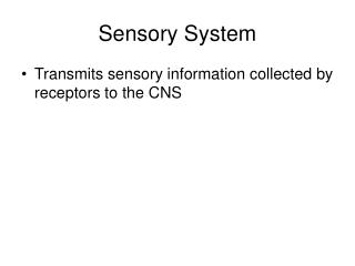
Test • What are the main sensory pathways (body and face), what function, and where it cross?
Anterolateral system (ALS): • Crude touch • Thermal sanitation • Pain • Anterior Trigeminothalamic pathway (Spinal Trigeminal Pathway): • Crude touch • Thermal sanitation • Pain Trunk, limbs, back of the head Face and from of the head
ALS Vs post. Colum pathways: (1) Generalized feeling of being touched but do not give precise localization, (2) Receptive fields are larger, (3) Fibres are smaller in diameter and more slowly conducting.
Peripheral Sensory and Motor Fibers: Groups, Diameters, and Conduction Velocities
Sensory dysfunction • Hypesthesia: reduced sensibility, • Paresthesia: numbness, tingling, and prickling, • Anesthesia loss of sensibility. • Allodynia: an innocuous stimulus will result in a perception of pain in the absence of a proper pain stimulus. • Hyperalgesia: is a heightened sensitivity to painful stimuli
Anterolateral System • spinothalamic fibers • spinomesencephalic fibers • spinoreticular fibers • spinobulbarfibers • spinohypothalamic fibers
Anterolateral System • Spinothalamic fibers project directly from the spinal cord to the ventral posterolateral (VPL) nucleus • Spinomesencephalic axons project to the periaqueductal area and to the tectum; the latter are spinotectal fibers. Play role in central modulaton of pain. • Spinoreticular tract: carried the emotional and arousal aspect of pain.
Figure 18-1 Summary of anterolateral system and anterior trigeminothalamic tract fibers conveying nondiscriminative tactile, thermal, and nociceptive inputs to the contralateral somatosensory cortex.
Figure 18-1 Summary of anterolateral system and anterior trigeminothalamic tract fibers conveying nondiscriminative tactile, thermal, and nociceptive inputs to the contralateral somatosensory cortex.
To summarize • If you step on a sharp object with your left foot, your spinothalamic tract enables you to realize “something sharp is puncturing the sole of my left foot”. • Your spinothalamic intralaminar projections and spinoreticular tract cause you to feel “ouch, that hurts!”; • And your spinomesencephalic tract leads to pain modulation, allowing you eventually to think “aah, that feels better”.
Peripheral Sensitization and Primary Hyperalgesia • Following an insult, • pain receptors become more sensitive • lower pain threshold • increases in firing rate to noxious stimulation. • So there will be increase in spontaneous activity in the Aδ and C fibers. • Although the mechanisms responsible for receptor sensitization are not completely known, chemicals released by the damaged skin or by products from plasma, or both, are thought to contribute to this phenomenon.
Peripheral Sensitization and Primary Hyperalgesia • As a result of this heightened sensitivity, the affected area is super-sensitive to painful stimuli and patients experience hyperalgesia. • Primary hyperalgesia: occurs in the region of damaged skin and is probably the result of receptor sensitization. • An example of primary hyperalgesia is the extreme sensitivity of sunburned skin, which results from sensitization of the skin pain endings by local tissue products from the burn-perhaps histamine, prostaglandins, and others
Central Sensitization and Secondary Hyperalgesia • Secondary hyperalgesia occurs in the skin bordering the damaged tissue. Although receptor sensitization may contribute to secondary hyperalgesia, there is likely to be a central (e.g., spinal) component as well. • There is hyper-activation of the cell in the posterior horn.
Central Sensitization and Secondary Hyperalgesia • This could be explain by: • increase in the receptive field size of the posterior horn neuron • an increased response of the cells to the application of suprathreshold stimuli, • This phenomenon is known as central sensitization, and it represents a potentiated state in which the system has been shifted from one functional level (normal) to another (sensitized) by a change in transcription.
Pain Receptors in Muscles, Joints, and Viscera • In addition to the cutaneous pain receptors, pain receptors in muscles, joints, and viscera have also been identified, which are also carried by III and IV afferent fibers type. • A pinprick exam usually activates Aδ fibers. • Dull, persistent ache that follows a muscle pull results from activation of C fibers.
Central Pathways • Aδ and C fibers enter the spinal cord via the lateral division of the posterior root entry zone. The fibers enter the posterolateral fasciculus (Lissauer tract) and bifurcate into ascending and descending branches
Summary of posterior horn laminae and their major sensory inputs (A) and major outputs (B).
Central Pathways • When Aδ fibers enter the posterolateral fasciculus and bifurcate, their branches travel rostrocaudally for three to five spinal levels. • The descending branches terminate on interneurons within the spinal gray that participate in segmental spinal reflexes.
The general somatotopic arrangement of the anterolateral system
Somatotopically Arrange DCT CST ALS ARM Trunk LEG
The ascending branches terminate on second-order neurons (tract cells) in lamina I of the posterior horn (Fig. 18-7A). These tract cells, in turn, project to the thalamus. • The great majority of their axons cross the midline of the spinal cord obliquely via the anterior (ventral) white commissure and ascend in the contralateral ALS. • A few ascend in the ipsilateral ALS. • The thalamic (third-order) neurons of these pathways are located mainly in the VPL, the posterior nucleus, and the intralaminar nuclei.
Blood supply to the anterolateral system in the spinal cord and medulla
Brown-Séquard syndrome • (1) contralateral loss of pain and thermal sensations over the body below the level of the lesion • (2) ipsilateral loss of discriminative tactile, vibratory, and position sense over the body below the level of the lesion • (3) ipsilateral paralysis of the leg or leg and arm, depending on the level of the hemisection.
Summary of the spinocervicothalamic tract that carries innocuous discriminative tactile, thermal, and nociceptive sensations.
Spinal Trigeminal Pathway: Anterior Trigeminothalamic Tract • Cranial nerves V, VII, IX, and X serve the cutaneous receptors of the face, the oral cavity. • The primary sensory fibers of these nerves have their cell bodies in the: • Trigeminal ganglion, • Geniculate ganglion of cranial nerve VII, • Superior ganglia of cranial nerves IX and X.
Anterior Trigeminothalamic Tract • Aδ and C fiber are found throughout the face and oral cavity. • The meninges are also supplied by fibers of the trigeminal ganglion cells that terminate in the spinal trigeminal nucleus.
Trigeminal sensory nucleus • Mesencephalic trigeminal nucleus • Propriception • Chief trigeminal nucleus • Fine touch • Spinal trigeminal nucleus • Pain, temp, crude touch
The spinal trigeminal nucleus • Divided into: • pars caudalis, • nondiscriminative touch, pain, and thermal sensations. • pars interpolaris, • pars oralis.
Pars caudalis cross section Trigeminal nerve distribution
Chronic pain Thalamic lesioning • Lateral thalamic lesions involve: VPL/VPM. • pain relief • S/E: loss of cutaneous and position sense in the affected limb, impaired motor function. • Medial thalamus lesion involve: the centromedian-parafascicular complex CM-PF, the central lateral nucleus, the medial dorsal nucleus. • Pain relief. • Less S/E
Chronic pain • Deep brain stimulation. • Stimulating electrodes centered in the somatosensory thalamus, the CM-PF complex, or the periventricular gray (PVG)-PAG activate neurons.
Test • What are the nucleus: • Mesencephalic trigeminal nucleus • Propriception • Chief trigeminal nucleus • Fine touch • Spinal trigeminal nucleus • Pain, temp, crude touch
Test • Draw a medulla cross section, and label the Lateral medullary (Wallenberg) syndrome.