Download
1 / 43
430 likes | 753 Vues
Chapter 32 Female Reproductive System. Overview: Female Reproductive System. Function to produce offspring and thereby ensure continuity of the genetic code It produces eggs, or female gametes, which each may unite with a male gamete to form the first cell of an offspring

E N D
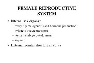
Overview: Female Reproductive System • Function • to produce offspring and thereby ensure continuity of the genetic code • It produces eggs, or female gametes, which each may unite with a male gamete to form the first cell of an offspring • It also can provide nutrition and protection to the offspring for up to several years after conception Slide
Overview: Female Reproductive System • Reproductive organs are classified as essential or accessory • Essential organs • gonads are the paired ovaries • Gametes are ova (eggs) produced by the ovaries • Accessory organs • Internal genitals • uterine tubes, uterus, and vagina • ducts that extend from the ovaries to the exterior • External genitals • the vulva • Mammary glands Slide
MRI Scan Slide
Overview: Female Reproductive System • Perineum • The perineum is the skin-covered region between the vaginal orifice and the rectum • This area may be torn during childbirth • extends anteriorly from symphysis pubis to coccyx posteriorly • lateral boundary is the ischial tuberosity on either side Slide
Ovaries • Location of the ovaries (ov = egg) • Nodular glands located on each side of the uterus, below and behind the uterine tubes • Large almonds • 3 g each • Ectopic pregnancy (ectop = displaced) • development of the fetus in a place other than the uterus Slide
Ovaries • Ovarian carcinoma Slide
Ovaries • Microscopic structure of the ovaries • Ovarian follicles • contain the developing female sex cells = oocyte (oo = egg) • Ovum • A developed oocyte released from the ovary Slide
Ovaries • Functions • Ovaries produce ova— the female gametes • Oogenesis— process that results in formation of a mature egg • Endocrine organs that secrete the female sex hormones- estrogens and progesterone Slide
Uterus • Structure • Size and shape • Pear-shaped, has two main parts— the cervix and the body • Wall of uterus is composed of three layers • inner endometrium • middle myometrium • Outer perimetrium Slide
Uterus Endometrial carcinoma Slide
Uterus • Cavities of uterus— cavities are small because of the thickness of the uterine walls • Internal os = apex of cervix • External os = opening of cervix Slide
Cadaver dissection showing uterine cavity and cervical canal, exposed by removal of parts of their posterior walls Slide
Uterus • Location of the uterus • Located in pelvic cavity between urinary bladder and rectum • Position of uterus is altered by age, pregnancy, and distention of related pelvic viscera • Descends, between birth and puberty, from the lower abdomen to the true pelvis • Begins to decrease in size at menopause Slide
Uterus • Position of the uterus • Body lies flexed over the bladder • Cervix points downward and backward, joining the vagina at a right angle • Several ligaments hold the uterus in place but allow some movement Slide
Uterus • Functions of the uterus • Part of reproductive tract, permits sperm to ascend toward uterine tubes • If conception occurs, offspring develops in the uterus • Embryo is supplied with nutrients by endometrial glands until the production of the placenta • Placenta is an organ that permits exchange of materials between mother’s blood and fetal blood but keeps the two circulations separate • Myometrial contractions occur during labor and help push the offspring out of mother’s body Slide
Uterus • If conception does not occur, outer layers of endometrium are shed during menstruation • Menstruation is a cyclical event that allows the endometrium to renew itself Slide
Endometrial (Menstrual) Cycle • 4 Phases over 28 days • Menses, menstrual period • Days 1-5 • Postmenstrual phase or preovulatory phase • Estrogenic or follicular phase • Days 6-13 • Ovulation • Days 14 • Premenstrual or postovulatory • Luteal phase or secretory phase or progesterone phase • Corpus luteum is secreting progesterone • Days 15-28 Slide
Endometrial (Menstrual) Cycle • Hypothalamus stimulates ovaries to make mature follicle • FSH- Follicle Stimulating Hormone • LH- Luteinizing Hormone • Maturing follicle releases estrogen and spikes (Day 13) • Hypothalamus responds with a burst of FSH and LH to release ovum • Ovulation (Day 14) • LH corpus luteum • Corpus luteum produces progesterone suppresses FSH & LH Slide
The rupture of a mature follicle on the surface of an ovary results in the release of an ovum into the pelvic cavity. The ovum released during ovulation is surrounded by a mass of cells. Slide
Uterine Tubes • Uterine tubes = fallopian tubes = oviducts • Structure of uterine tubes • Consist of mucous, smooth muscle, and serous lining • Tubal mucosa is continuous with vagina and uterus can become infected with organisms introduced into the vagina • Function of the uterine tubes • Serve as transport channels for ova and as the site of fertilization Slide
Tubal ligation Slide
Vagina • Vagina is a tubular organ located between the rectum, the urethra, and the bladder • Structure of the vagina • Collapsible tube capable of distention • 7 or 8 cm long (3 inches) • composed of smooth muscle • lined with mucous membrane arranged in rugae • Hymen— a mucous membrane that typically forms a border around the vagina in young premenstrual girls Slide
Vagina • Functions of the vagina • Lining of the vagina lubricates and stimulates the penis during sexual intercourse and acts as a receptacle for semen • Lower portion of the birth canal • Transports tissue and blood shed during menstruation to the exterior Slide
Vulva • The vulva consists of the female external genitals: • mons pubis (pad of fat over symphysis pubis) • labia majora and minora • Clitoris • urinary meatus (urethral orifice) • vaginal orifice • greater vestibular gland Slide
Vulva • Functions of the vulva • The mons pubis and labia protect the clitoris and vestibule • The clitoris contains sensory receptors that send information to the sexual response area of the brain • The vaginal orifice is the boundary between the internal and external genitals Slide
Breasts • Location and size • The breasts lie over the pectoral muscles • Estrogens and progesterone control breast development • 15-20 lobes • Breast size is determined by the amount of fat around glandular tissue not related to functional ability • Areola- becomes darker during pregnancy Slide
Breasts • Function of the breasts • Lactation • Mechanism of lactation • Ovarian hormones make breasts structurally ready to produce milk • Estrogen promotes duct development • Progesterone promotes development of alveoli, the secreting cells • Shedding of placenta decrease of estrogens stimulates prolactin stimulates lactation • Suckling also stimulates lactation • Secretion starts about 3-4 days after delivery • Oxytocin is released to facilitate bonding between mother and child Slide
Breasts • Lactation can provide nutrient-rich milk to offspring for up to several years from birth • Some advantages are the following: • Nutrients • Passive immunity from antibodies present in colostrum and milk • Emotional bonding between mother and child Slide
Female Reproductive Cycles • The female reproductive system has many cyclical changes that start with the beginning of menses • Ovarian cycle— ovaries from birth contain oocytes in primary follicles • at the beginning of menstruation each month, several of the oocytes resume meiosis • meiosis will stop again just before the cell is released during ovulation • Menstrual cycle (endometrial cycle) is divided into four phases: • Menses • Postmenstrual phase • Ovulation • Premenstrual phase Slide
Female Reproductive Cycles • Control of female reproductive cycles • Hormones control cyclical changes • Cyclical changes in ovaries result from changes in gonadotropins (FSH and LH) secreted by pituitary gland • Cyclical changes in uterus are caused by changes in estrogens and progesterone • Low levels of FSH and LH cause regression of the corpus luteum if pregnancy does not occur • this causes a decrease in estrogen and progesterone, which triggers endometrial sloughing of the menstrual phase • Control of cyclical changes in gonadotropin secretion is caused by positive and negative feedback mechanisms and involves estrogens, progesterone, and the hypothalamus’s secretion of releasing hormones Slide
Female Reproductive Cycles • Importance of the female reproductive cycles • Ovarian cycle’s primary function is to produce an ovum at regular intervals • Secondary function is to regulate endometrial cycle through estrogen and progesterone • Function of endometrial cycle is to make uterus suitable for implantation of a new offspring • Cyclical nature of the reproductive system and the fact that fertilization will occur within 24 hours after ovulation mean that a woman is only fertile a few days of each month • Menstrual flow begins at puberty, and menstrual cycle continues for about three decades Slide
The Big Picture: The Female Reproductive System and the Whole Body • The female reproductive system shares a special relationship with the following: • The urinary system because of their close proximity and because they share the vulva • The skeletal muscles in the perineum • The integumentary system because breasts are actually modifications of the skin Slide
Pictures of gynecological system pathology • http://www.uphs.upenn.edu/path/web_docs/p200/GYN200/GYN95.html Slide
Hydrops fetalis- abnormal accumulation of fluid in fetal compartments Slide
Placenta percreta • Placenta attaches itself too firmly to the uterus, going through the myometrium and serosa (ruptures the uterus) • 1: 2500 pregnancies Slide