Wrist Sonography
Wrist Sonography. Caitlin Gardiner. Preparation. Have patient sitting on a chair across with exposing their anterior wrist and resting their hand on the table with an absorbent sheet beneath Select a high-frequency linear probe with a hockey stick probe if available


Wrist Sonography
E N D
Presentation Transcript
Wrist Sonography Caitlin Gardiner
Preparation • Have patient sitting on a chair across with exposing their anterior wrist and resting their hand on the table with an absorbent sheet beneath • Select a high-frequency linear probe with a hockey stick probe if available • Ideally a thick coupling gel is used due to the hand contours
Purpose of Ultrasound • Chronic and Acute muscular, ligament and tendon damage • Joint effusion • Bursitis • Haematoma • Ganglions/ other solid or cystic lesions • Bony surface • Dynamic assessment of tendons and relationships
Volvar Aspect of Wrist • Proximal Carpal Tunnel
Volvar Aspect of Wrist • DistalCarpal Tunnel
Assessing Flexor Tendons • Start transverse, scan to distal insertion, turn long when assessing dynamic motion. • Tear due to direct or non-direct trauma • Tear location need to be assess as well as the retraction of the tendon ends • Assess for typical fibrillar echotexture • Proximal end of a tear will show retracted tendon (swollen, irregular and hypoechoic) which will not move on dynamic evaluation • Most commonly tears occur of the profundus tendon just proximal to its insertion • In entrapment • Hypoechoic halo surrounding the tendon sheath will be more distinct
Assessing the Retinacula • Powerful traction can cause tears • Dislocation of the tendon can be found medially, close to the extensor digitorum minimi or medial to the ulnar head • In entrapment conditions • Volvar bulging secondary to increases in intracanal pressure • Measure, at the distal end of the carpel tunnel, the distance between an arbitrary line from a) the hook of the hamate to the tubercle of the trapezium to b) the retinaculum and ensure the distance is not more than 4mm
Nerves of the Volvar Aspect • Median Nerve • Enlarged in Carpel Tunnel Syndrome • Can be easily tracked up the forearm • Image in transverse and measure 2D volume at widest point • Image in longitudinal • In Carpal Tunnel syndrome/entrapment, • Swollen at proximal portion (>10-12mm²) • Decrease in overall echogenicity and normal fascicular pattern • Increase in vascularity in severe cases
Nerves of the Volvar Aspect • Radial Nerve • Clinically significant if inflamed as it crosses the first extensor compartment to reach the dorsal aspect of the wrist • Ulnar Nerve • Proximal: Lies within Guyon’s canal between the ulnar artery and the pisiform • Distal: Divides into a superficial and deep motor branch. The deep branch can be damaged by hook of the mate by compression
Nerve Tumours • Mostly affect median nerve and ulna nerve • Compression can cause tingling • Neurinomas • Embedded inside the nerve and never fascicles are seen transverse within them • Easily surgically removed • Neurofibromas • Arise at the periphery of the nerve and grow eccentrically
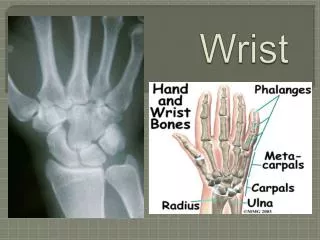
Transverse Dorsal Aspect • First position probe in transverse on distal forearm so the radius and ultra are obtained. • Move distally across the radio-carpal joint (where two bones become three; the scaphoid, triquetral and lunate). • Note any ganglion as a poorly reflective fluid collection.
Transverse Dorsal Aspect • Note six compartments
Assessing Extensor Tendons • Tears often occur as a result of rheumatoid tendosynovitis, causing friction between tendons and bon protuberances (Ulnar head and Lister’s tubercle) • Most commonly affected are the extensor digiti minimi and the extensor pollicis longus
Masses of the Wrist • Describe • Location: subcutaneous, subfascial plane or adherent to bone plane (measure distance to the skin for biopsy/surgery) • Borders: Regular, irregular or dendritic • Vascularity • Relationship to surrounding structures • Dynamic Behaviour (moves with tendons, compression etc) • Ganglia appear as anechoic structures with internal septa and has a fibrous wall and lacks a true synovial lining. They most commonly occur in the dorsal aspect of the wrist. Typically painless, firm masses
Other Lesions • Subcutaneous and muscle haematoma appear as fluid collections • Abscess (following penetrating injury) appears as a poorly defined heterogeneous mass with surrounding hyperaemia • Post-traumatic Intra-articular effusion can be visualised as a collection filling the joint space and the articular synovial recesses • Radiolucent foreign bodies can be detect on US (though x-ray shows radio-opaque bodies)
References • Beggs I, Bianchi S, Bueno A et al. Musculoskeletal Technical Guidelines: Wrist. European Society of Musculoskeletal Radiology. • Bianchi S and Matinoli C, 2007. Ultrasound of the Musculoskeletal System. Springer, Geneva. • McNally E, 2005. Practical Musculoskeletal Ultrasound. Elsevier Churchill Livingstone, Philadelphia.