Download

1 / 10
100 likes | 399 Vues
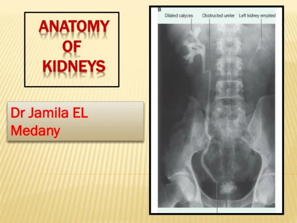
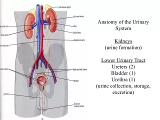
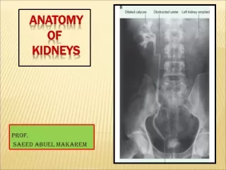
Surgical Anatomy of the surgical anatomy of the Retroperitoneum , Adrenals, Kidneys, and Ureters. There is no greater aid to surgical expertise than an intimate knowledge of anatomy. For the urologist, the areas of greatest importance are the retroperitoneum and pelvis.

E N D
Surgical Anatomy of thesurgical anatomy of the Retroperitoneum, Adrenals,Kidneys, and Ureters
There is no greater aid to surgical expertise than an intimate knowledge of anatomy. For the urologist, the areas of greatest importance are the retroperitoneum and pelvis.
l The lower ribs are in intimate contact with the kidneys and adrenal glands. Injury to the lower ribs suggests injury to retroperitoneal structures. l The renal artery lies posterior to the renal vein, but this relationship is reversed when the aorta and inferior vena cava divide into the common iliac vessels. Here, the common iliac arteries are anterior to the common iliac veins.
Psoas and Iliacus Muscles The psoas major muscle originates on the 12th thoracic through the 5th lumbar vertebrae . A smaller psoas minor is identifiable in about one half of the population and resides medial to the psoas major. The psoas muscle(s) is covered by the psoas fascia. In close proximity to the psoas muscle is the iliacus muscle, which attaches to the inner aspect of the iliac pelvic wing. As the iliacus progresses caudally it joins with the psoas muscle to form the iliopsoas muscle. This combined muscle then joins to the lesser trochanter of the femur and controls flexion of the hip.
Externaloblique Lower eight ribs Lateral lip of iliac crest, aponeurosis ending in midline raphe Compress abdominal contents, flexion of the trunk Internal oblique Lumbodorsal fascia, iliac crest Lower 4 ribs, aponeurosis ending in linea alba Compress abdominal contents, flexion of the trunk • Transversus • abdominis • Lumbodorsal • fascia, medial • lip of iliac crest • Aponeurosis • ending in • linea alba • Compress • abdominal • contents