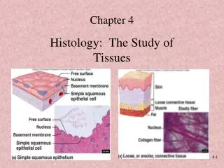
Chapter 12: Neural Tissue
Chapter 12: Neural Tissue. Functions of the CNS . Are to process and coordinate: sensory data: from inside and outside body motor commands: control activities of peripheral organs (e.g., skeletal muscles) higher functions of brain: intelligence, memory, learning, emotion. Neural Tissue.


Chapter 12: Neural Tissue
E N D
Presentation Transcript
Functions of the CNS • Are to process and coordinate: • sensory data: • from inside and outside body • motor commands: • control activities of peripheral organs (e.g., skeletal muscles) • higher functions of brain: • intelligence, memory, learning, emotion
Neural Tissue • Contains 2 kinds of cells: • neurons: • cells that send and receive signals • neuroglia (glial cells): • cells that support and protect neurons
Organs of the Nervous System • Brain and spinal cord • Sensory receptors of sense organs (eyes, ears, etc.) • Nerves connect nervous system with other systems
Anatomical Divisions of the Nervous System • Central nervous system (CNS) • The Brain and Spinal Cord • Peripheral nervous system (PNS) • Nerves and ganglia out side of the CNS. • Includes the “Motor” and “Sensory” divisions
Functions of the PNS • Deliver sensory information to the CNS • Carry motor commands to peripheral tissues and systems
Nerves • Also called peripheral nerves: • bundles of axons with connective tissues and blood vessels • carry sensory information and motor commands in PNS: • cranial nerves—connect to brain • spinal nerves—attach to spinal cord
Functional Divisions of the PNS • Afferent division: • carries sensory information • from PNS sensory receptors to CNS • Efferent division: • carries motor commands • from CNS to PNS muscles and glands
Receptors and Effectors • Receptors: • detect changes or respond to stimuli • neurons and specialized cells • complex sensory organs (e.g., eyes, ears) • Effectors: • respond to efferent signals • cells and organs
The Efferent Division of the PNS • Somatic nervous system (SNS) • Autonomic nervous system (ANS)
The Somatic Nervous System (SNS) • Controls skeletal muscle contractions: • voluntary muscle contractions • involuntary muscle contractions (reflexes)
The Autonomic Nervous System(ANS) • Controls subconscious actions: • contractions of smooth muscle and cardiac muscle • glandular secretions
Divisions of the ANS • Sympathetic division: • has a stimulating effect • Parasympathetic division: • has a relaxing effect
The Structure of Neurons PLAY Neurophysiology: Neuron Structure Figure 12–1
The Multipolar Neuron • Common in the CNS: • cell body (soma) • short, branched dendrites • long, single axon
Major Organelles of the Cell Body • Large nucleus and nucleolus • Cytoplasm (perikaryon) • Mitochondria (produce energy) • RER and ribosomes (produce neurotransmitters) • Cytoskeleton
The Cytoskeleton • Neurofilaments and neurotubules: • in place of microfilaments and microtubules • Neurofibrils: • bundles of neurofilaments • support dendrites and axon
Nissl Bodies • Dense areas of RER and ribosomes • Make neural tissue appear gray (gray matter)
Dendrites • Highly branched • Dendritic spines: • many fine processes • receive information from other neurons • 80–90% of neuron surface area
Structures of the Axon • Axoplasm: • cytoplasm of axon • contains neurotubules, neurofibrils, enzymes, organelles • Axolemma: • specialized cell membrane • covers the axoplasm
Structures of the Axon • Axon hillock: • thick section of cell body • attaches to initial segment • Initial segment: • attaches to axon hillock
Structures of the Axon (3 of 3) • Collaterals: • branches of a single axon • Telodendria: • fine extensions of distal axon • Synaptic terminals: • tips of axon
The Synapse PLAY Neurophysiology: Synapse Figure 12–2
Neurotransmitters • Are chemical messengers • Are released at presynaptic membrane • Affect receptors of postsynaptic membrane • Are broken down by enzymes • Are reassembled at synaptic knob
Recycling Neurotransmitters • Axoplasmic transport: • neurotubules within the axon • transport raw materials • between cell body and synaptic knob • powered by mitochondria and kinesins
Types of Synapses • Neuromuscular junction: • synapse between neuron and muscle • Neuroglandular junction: • a synapse between neuron and gland
4 Structural Classifications of Neurons • Anaxonic neurons: • found in brain and sense organs • Bipolar neurons: • found in special sensory organs (sight, smell, hearing)
4 Structural Classifications of Neurons • Unipolar neurons: • found in sensory neurons of PNS • Multipolar neurons: • common in the CNS • include all skeletal muscle motor neurons
Anaxonic Neurons • Small • All cell processes look alike Figure 12–3 (1 of 4)
Bipolar Neurons • Are small • 1 dendrite, 1 axon Figure 12–3 (2 of 4)
Unipolar Neurons • Are very long axons • Fused dendrites and axon • Cell body to 1 side Figure 12–3 (3 of 4)
Multipolar Neurons • Have very long axons • Multiple dendrites, 1 axon Figure 12–3 (4 of 4)
3 Functional Classifications of Neurons • Sensory neurons: • afferent neurons of PNS • Motor neurons: • efferent neurons of PNS • Interneurons: • association neurons
3 Types of Sensory Receptors • Interoceptors: • monitor internal systems (digestive, respiratory, cardiovascular, urinary, reproductive) • internal senses (taste, deep pressure, pain) • Exteroceptors: • external senses (touch, temperature, pressure) • distance senses (sight, smell, hearing) • Proprioceptors: • monitor position and movement (skeletal muscles and joints)
Motor Neurons • Carry instructions from CNS to peripheral effectors • Via efferent fibers (axons)
2 Major Efferent Systems • Somatic nervous system (SNS): • includes all somatic motor neurons that innervate skeletal muscles • Autonomic (visceral) nervous system (ANS): • visceral motor neurons innervate all other peripheral effectors: • e.g., smooth muscle, cardiac muscle, glands, adipose tissue
2 Groups of Efferent Axons • Signals from CNS motor neurons to visceral effectors pass synapses at autonomic ganglia dividing axons into: • preganglionic fibers • postganglionic fibers
Interneurons • Most are located in brain, spinal cord, and autonomic ganglia: • between sensory and motor neurons • Are responsible for: • distribution of sensory information • coordination of motor activity • Are involved in higher functions: • memory, planning, learning
Neuroglia of the Central Nervous System Figure 12–4
4 Types of Neuroglia in the CNS • Ependymal cells: • highly branched processes • contact neuroglia directly • Astrocytes: • large cell bodies • many processes
4 Types of Neuroglia in the CNS • Oligodendrocytes: • smaller cell bodies • fewer processes • Microglia: • small • many fine-branched processes
Ependymal Cells • Form epithelium called ependyma • Line central canal of spinal cord and ventricles of brain: • secrete cerebrospinal fluid (CSF) • have cilia or microvilli that circulate CSF • monitor CSF • contain stem cells for repair
Astrocytes • Maintain blood–brain barrier (isolates CNS) • Create 3-dimensional framework for CNS • Repair damaged neural tissue • Guide neuron development • Control interstitial environment
Oligodendrocytes • Processes contact other neuron cell bodies • Wrap around axons to form myelin sheaths
Myelination • Increases speed of action potentials • Myelin insulates myelinated axons • Makes nerves appear white
Nodes and Internodes • Internodes: • myelinated segments of axon • Nodes: • also called nodes of Ranvier • gaps between internodes • where axons may branch
White Matter and Gray Matter • White matter: • regions of CNS with many myelinated nerves • Gray matter: • unmyelinated areas of CNS
Microglia • Migrate through neural tissue • Clean up cellular debris, waste products, and pathogens
Ganglia • Masses of neuron cell bodies • Surrounded by neuroglia • Found in the PNS