Glycolysis
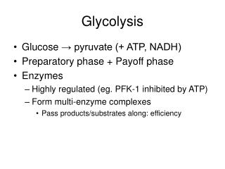
Glycolysis. UNIT II: Intermediary Metabolism. Overview. In cells reactions rarely occur in isolation, but rather organized into multi-step sequences called pathways, such as glycolysis In a pathway, product of one reaction serves as substrate of subsequent reaction

Glycolysis
E N D
Presentation Transcript
Glycolysis UNIT II: Intermediary Metabolism
Overview • In cells reactions rarely occur in isolation, but rather organized into multi-step sequences called pathways, such as glycolysis • In a pathway, product of one reaction serves as substrate of subsequent reaction • Different pathways can also intersect, forming an integrated and purposeful network of chemical reactions. These are collectively called metabolism, which is the sum of all chemical changes occurring in a cell, a tissue or the body. • Most pathways can be classified as either catabolic (degradative) or anabolic (synthetic). • Catabolic reactions break down complex molecules e.g., proteins, polysacch’s & lipids, to a few simple molecules e.g., CO2, NH3 & H2O • Anabolic pathway form complex end products from simple precursors e.g., synthesis of the polysacch, glycogen, from glucose
Figure 8.1 Glycolysis, an example of a metabolic pathway.
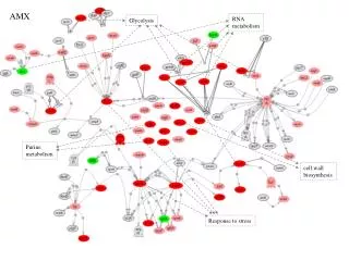
Metabolic map - It is convenient to investigate metabolism by examining its component pathways • Each pathway is multienzyme sequences, and each enz in turn may exhibit important catalytic or regulatory feature • Metabolic map is useful in tracing connections b/w pathways, visualizing purposeful movement of metabolic intermediates, and picturing the effect on flow of intermediates if a pathway is blocked e.g., by a drug or inherited deficiency of an enz
Figure 8.2. Important reactions of intermediary metabolism. Several important pathways to be discussed in later chapters are highlighted. Curved reaction arrows ( ) indicate forward and reverse reactions that are catalyzed by different enzymes. The straight arrows ( ) indicate forward and reverse reactions that are catalyzed by the same enzyme. Key: Blue text = intermediates of carbohydrate metabolism; brown text = intermediates of lipid metabolism; green text = intermediates of protein metabolism.
B. Catabolic pathways • Catabolic reactions serve to capture chemical energy in the form of ATP from degradation of energy-rich fuel molecules • Catabolism also allows molecules in diet (or nutrient molecules stored in cells) to be converted to building blocks needed for synthesis of complex molecules • Energy generation by degradation of complex molecules occurs in 3 stages: 1. Hydrolysis of complex molecules: complex molecules are broken down into their component building blocks. E.g., proteins aa’s, polysacch’s monosacch’s & triglycerides free fatty acids and glycerol
2. Conversion of building blocks to simple intermediates: diverse building blocks further degraded to acetyl CoA and a few other simple molecules. Some energy is captured as ATP, but amount is small compared with that produced during 3rd stage 3. Oxidation of acetyl CoA: TCA cycle is the final common pathway in oxidation of fuel molecules such as acetyl CoA. Large amounts of ATP are generated as e’s flow from NADH & FADH2 to O2 via oxphos
C. Anabolic pathway • Anabolic reactions combine small molecules, e.g., aa’s to form complex molecules, such as proteins • Anabolic reactions require energy, which is generally provided by breakdown of ATP to ADP + Pi • Anabolic reactions often involve chemical reductions in which reducing power is most frequently provided by e-donor NADPH • Note that catabolism is a convergent process i.e., a wide variety of molecules are transformed into a few common end products • By contrast, anabolism is a divergent process i.e., a few biosynthetic precursors form a wide variety of polymeric or complex products
II. Regulation of metabolism • Pathways of metabolism must be coordinated so that production of energy or synthesis of end products meets needs of cell • Individual cells do not function in isolation but, rather, are part of a community of interacting tissues • Thus, a sophisticated communication system has evolved to coordinate functions of the body • Regulatory signals that inform an individual cell of the metabolic state of the body as a whole include hormones, neurotransmitters, and the availability of nutrients. These, in turn, influence signals generated within the cell
Signals from within the cell (intracellular) • Rate of a metabolic pathway can respond to regulatory signals that arise from within cell • E.g., rate of a pathway may be influenced by availability of substrates, product inhibition, or alterations in levels of allosteric activators or inhibitors • These intracellular signals typically elicit rapid responses, and are important for moment-to-moment regulation of metabolism
B. Communication between cells (intercellular) • Ability to respond to extracellular signals is essential for survival & development of all organisms • Signaling b/w cells provides for long-range integration of metabolism, & usually results in a response that is slower than is seen with signals that originates within the cell • Communication b/w cells can be mediated by surface-to-surface contact &, in some tissues, by formation of gap junctions, allowing direct communication b/w cytoplasms of adjacent cells • However, for energy metabolism, the most important route of communication is chemical signaling e.g., by blood-borne hormones or by neurotransmitters
C. Second messenger systems • Hormones or neurotransmitters can be thought of as signals, & a receptor as a signal detector. Each component serves as a link in the communication b/w extracellular events & chemical changes within the cell • Many receptors signal their recognition of a bound ligand by initiating a series of reactions that ultimately result in a specific intracellular response • “second messenger” molecules, so named as they intervene b/w original messenger (neurotransmitter or hormone) & the ultimate effect on cell, are part of the cascade of events that translates hormone or neurotransmitter binding into a cellular response • Two of the most widely recognized 2nd messenger systems are the calcium/phosphatidylinositol system & adenylyl cyclase system, which is particularly important in regulating pathways of intermediary metabolism
Figure 8.5 Some commonly used mechanisms for transmission of regulatory signals between cells.
D. Adenylyl cyclase • Recognition of a chemical signal by some memb receptors, such as β- & α2-adrenergic receptors, triggers either an increase or a decrease in the activity of adenylyl cyclase. • This is a memb-bound enz that converts ATP to 3`,5`-adenosine monophosphate (a.k.a cyclic AMP or cAMP) • Chemical signals are most often hormones or neurotransmitters, each of which binds to a unique type of memb receptor • Therefore, tissues that respond to more than one chemical signal must have several different receptors, each of which can be linked to adenylyl cyclase Note: certain toxins, as that produced by Vibrio cholera, can also activate the adenylyl cyclase cascade, with potentially disastrous consequences - These receptors are characterized by an extracellular ligand-binding region, 7 transmembrane helices, & an intracellular domain that interacts with G-proteins
Figure 8.6 Structure of a typical membrane receptor.
GTP-dependent regulatory proteins: • Effect of activated occupied receptor on 2nd messenger formation is not direct, it is mediated by specialized trimeric proteins in the CM. • These, referred to as G-proteins because they bind guanosine nucleotides (GTP & GDP), form a link in the chain of communication b/w receptor & adenylyl cyclase • The inactive form of G-protein binds to GDP • The activated receptor interacts with G-proteins, triggering an exchange of GTP for GDP. • The timeric G-proteins then dissociates into an α subunit & a βγ dimer. • The GTP-bound form of the α subunit moves from the receptor to adenylyl cyclase, which is thereby activated
Many molecules of active G-protein are formed by one activated receptor Note: ability of a hormone or neurotransmitter to stimulate or inhibit adenylyl cyclase depends on the type of G-protein that is linked to the receptor. One family of G-proteins, designated Gs, is specific for stimulation of adenylyl cyclase; another family, Gi inhibition of the enz. - Actions of G-protein-GTP complex are short-lived because the G-protein has an inherent GTPase activity rapid hydrolysis of GTP to GDP inactivation of G-protein
Figure 8.7. The recognition of chemical signals by certain membrane receptors triggers an increase (or, less often, a decrease) in the activity of adenylyl cyclase.
2. Protein kinases: • Next link in cAMP 2nd-messenger system is activation by cAMP of a family of enz’s = cAMP-dependent protein kinases, e.g., protein kinase A. • cAMP activates protein kinase A by binding to its two regulatory subunits, causing release of active catalytic subunits. • The active subunits catalyze the transfer of P from ATP to specific ser or thr residues of protein substrates • Phosphorylated proteins may act directly on cell’s ion channels, or may become activated or inhibited enz’s • Protein kinase A can also phosphorylate specific proteins that bind to promoter regions of DNA increased expression of specific genes • Note: not all protein kinases respond to cAMP; there are several types of protein kinases that are not cAMP-dependent, e.g., protein kinase C
3. Dephosphorylation of proteins: • P groups added to proteins by protein kinases are removed by protein phosphatases, enz’s that hydrolytically cleave phosphate esters • This ensures that changes in enzymatic activity induced by protein phosphorylation are not permanent
4. Hydrolysis of cAMP: • cAMP is rapidly hydrolyzed to 5`-AMP by cAMP phosphodiesterase, one of a family of enz’s that cleave cyclic 3`,5`-phosphodiester bond • 5`-AMP is not an intracellular signaling molecule. Thus, effects of neurotransmitter or hormone-mediated increases of cAMP are rapidly terminated if the extracellular signal is removed Note: phosphodiesterase is inhibited by methylxanthine derivatives, such as theophylline & caffeine
III. Overview of glycolysis • Glycolytic pathway is employed by all tissues for the breakdown of glucose to provide energy (in form of ATP) and intermediates for other metabolic pathways • Glycolysis is at the hub of CHO metabolism because virtually all sugars, whether arising from diet or from catabolic reactions in the body, can ultimately be converted to glucose • Pyruvate is the end product of glycolysis in cells with mitochondria & an adequate supply of oxygen • This series of 10 reactions is called aerobic glycolysis because oxygen is required to reoxidize NADH formed during oxidation of glyceraldehyde-3-P
Aerobic glycolysis sets the stage for the oxidative decarboxylation of pyruvate to acetyl CoA, a major fuel of TCA cycle • Alternatively, glucose can be converted to pyruvate, which is reduced by NADH lactate. This is called anaerobic glycolysis because it can occur without participation of oxygen. • Anaerobic glycolysis allows the continued production of ATP in tissues that lack mitochondria (e.g., RBCs) or in cells deprived of sufficient oxygen
Figure 8.9. A. Glycolysis shown as one of the essential pathways of energy metabolism. B. Reactions of aerobic glycolysis. C. Reactions of anaerobic glycolysis.
IV. Transport of glucose into cells • Glucose cannot diffuse directly into cells, but enters by one of two transport mechanisms: a Na+-independent, facilitated diffusion transport system, or a Na+-monosacch co-transporter system A. Na+-independent facilitated diffusion transport • This system is mediated by a family of at least 14 glucose transporters in CMs. They are = GLUT-1 to GLUT-14 (glucose transporter isoform 1-14) • These transporters exist in membrane in two conformational states. Extracellular glucose binds to the transporter, which then alters its conformation, transporting glucose across the CM
Figure 8.10 Schematic representation of the facilitated transport of glucose through a cell membrane.
1. Tissue specificity of GLUT gene expression: - Glucose transporters display a tissue-specific pattern of expression e.g., GLUT-3 is the primary glucose transporter in neurons. GLUT-1 is abundant in erythrocytes & brain, but is low in adult muscle, whereas GLUT-4 is abundant in adipose tissue & skeletal muscle Note: number of GLUT-4 transporters active in these tissues is increased by insulin. The other GLUT isoforms also have tissue-specific distributions
2. Specialized functions of GLUT isoforms: • In facilitated diffusion, glucose movement follows a conc. gradient, i.e., from a high gluc conc. to a lower one. E.g., GLUT-1, -3, -4 are primarily involved in gluc uptake from blood • In contrast, GLUT-2 which is found in liver, kidney, and β-cells of pancreas, can either transport gluc into these cells when blood gluc levels are high, or transport gluc from cells to blood when blood glucose levels are low (e.g., during fasting) • GLUT-5 is unusual in that it is the primary transporter for fructose (instead of gluc) in the small intestine & testes • GLUT-7 which is expressed in liver & other gluconeogenic tissues, mediates glucose flux across endoplasmic reticulum memb.
B. Na+-monosaccharide cotransporter system • This is an energy-requiring process that transports glucose “against” a conc. gradient i.e., from low gluc conc’s outside the cell to higher conc’s within cell • This system is a carrier-mediated process in which movement of glucose is coupled to the conc gradient of Na+, which is transported into cell at the same time. • This type of transport occurs in epithelial cells of intestine, renal tubules, and choroid plexus
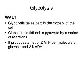
V. Reactions of glycolysis • Conversion of gluc to pyruvate occurs in 2 stages. The 1st five reactions correspond to an energy investment phase in which phosphorylated forms of intermediates are synthesized at the expense of ATP • The subsequent reactions of glycolysis constitute an energy generation phase in which a net of 2 molecules of ATP are formed by substrate level phosphorylation per gluc molecule metabolized Note: 2 molecules of NADH are formed when pyruvate is produced (aerobic glycolysis), whereas NADH is converted to NAD+ when lactate is produced (anaerobic glycolysis)
Figure 8.11 Two phases of aerobic glycolysis.
Phosphorylation of glucose • Phosphorylated sugar molecules do not readily penetrate CMs, because no specific transmemb carriers for these cpds, & they are too polar to diffuse through the CM • The irreversible phosphorylation of gluc, therefore, effectively traps the sugar as cytosolic G-6-P, thus committing it to further metabolism in the cell • Mammals have several isozymes of the enz hexokinase that catalyze the phosphrylation of gluc to G-6-P
Figure 8.12 Energy investment phase: phosphorylation of glucose.
1. Hexokinase: • In most tissues, phosphorylation of gluc is catalyzed by hexokinase, one of 3 regulatory enz’s of glycolysis (phosphofructokinase I & pyruvate kinase are the other two) • Hexokinase has broad substrate specificity and is able to phosphorylate several hexoses in addition to gluc. • Hexokinase is inhibited by the reaction product, G-6-P, which accumulates when further metabolism of this hexose-P is reduced • Hexokinase has a low Km (&, therefore, a high affinity) for gluc. This permits efficient phosphorylation & subsequent metabolism of gluc even when tissue conc’s of gluc are low • Hexokinase, however, has a low Vmax for gluc &, therefore, cannot sequester (trap) cellular phosphate in the form of phosphorylated hexoses, or phosphorylate more sugars than the cell can use
2. Glucokinase: • In liver parenchymal cells & islet cells of the pancreas, glucokinase (also called hexokinase D, or type IV) is the predominant enz responsible for the phosphorylation of gluc • In β-cells, glucokinase functions as gluc sensor, determining the threshold for insulin secretion • In liver, the enz facilitates gluc phosphorylation during hyperglycemia Note: despite its misleading name “glucokinase” the sugar specificity of the enz is similar to that of other hexokinase isozymes
a. Kinetics: • Glucokinase differs from hexokinase in several important properties • E.g., it has much higher Km, requiring a higher gluc conc for half-saturation. Thus glucokinase functions only when intracellular conc of gluc in hepatocyte is elevated, e.g., during the brief period following consumption of a CHO-rich meal, when high levels of gluc are delivered to the liver via the portal vein • Glucokinase has a high Vmax, allowing the liver to effectively remove the flood of gluc delivered by the portal blood. This prevents large amounts of gluc from entering systemic circulation following a CHO-rich meal, & thus minimizes hyperglycemia during the absorptive period Note: GLUT-2 insures that blood gluc equilibrates rapidly across the memb of the hepatocyte
Figure 8.13 Effect of glucose concentration on the rate of phosphorylation catalyzed by hexokinase and glucokinase.
b. Regulation by fructose 6-phosphate and glucose: • Glucokinase activity is not allosterically inhibited by G-6-P as are other hexokinases, but rather is indirectly inhibited by F-6-P (which is in equil. with G-6-P), & is stimulated indirectly by glucose via the following mechanism: • A glucokinase regulatory protein exists in the nucleus of hepatocytes. • In presence of F-6-P, glucokinase is translocated into nucleus & binds tightly to regulatory protein, thus rendering enz inactive • When gluc levels in the blood (& also in the hepatocyte, as a result of GLUT-2) increase, the gluc causes the release of glucokinase from the regulatory protein, & the enz enters the cytosol where it phosphorylates gluc to G-6-P • As free gluc levels fall, F-6-P causes glucokinase to translocate back into nucleus & bind to regulatory protein, thus inhibiting enz’s activity
Figure 8.14 Regulation of glucokinase activity by glucokinase regulatory protein.
c. Regulation by insulin: - Glucokinase activity in hepatocytes is also increased by insulin - as blood gluc levels rise following a meal, β-cells of pancreas are stimulated to release insulin into portal circulation Note: ~ ½ of the newly secreted insulin is extracted by liver during the 1st pass through that organ. Therefore, liver is exposed to twice as much insulin as is found in systemic circulation - insulin also promotes transcription of glucokinase gene, resulting in an increase in liver enz protein &, therefore, of total glucokinase activity. Note: the absence of insulin in patients with diabetes causes a deficiency in hepatic glucokinase. This contributes to an inability of patient to efficiently decrease blood glucose levels
B. Isomerization of glucose-6-phosphate • Isomerization of G-6-P to F-6-P is catalyzed by phosphoglucose isomerase. Reaction is readily reversible & is not a rate-limiting or regulated step Figure 8.15 Isomerization of glucose 6-phosphate to fructose 6-phosphate.
C. Phosphorylation of fructose 6-phosphate • The irreversible phosphorylation reaction catalyzed by phosphofructokinase-1 (PFK-1) is the most important control point & the rate-limiting step of glycolysis • PFK-1 is controlled by available conc’s of the substrates ATP & F-6-P, & by the following regulatory substances: 1. Regulation by energy levels within the cell: • PFK-1 is inhibited allosterically by elevated levels of ATP, which act as an “energy-rich” signal indicating an abundance of high-energy cpds. • Elevated levels of citrate, an intermediate in the TCA cycle, also inhibit PFK-1. Conversely, PFK-1 is activated allosterically by high conc’s of AMP, which signal that the cells’ energy stores are depleted
2. Regulation by fructose 2,6-bisphosphate • F-2,6-bisP is the most potent activator of PFK-1. This cpd also acts as an inhibitor of fructose 1,6-bisphosphatase • The reciprocal actions of F-2,6-bisP on glycolysis & gluconeogenesis ensure that both pathways are not fully active at the same time Note: this would result in a “futile cycle” in which glucose would be converted to pyruvate followed by re-synthesis of glucose from pyruvate • F-2,6-bisP is formed PFK-2, an enz different than PFK-1. F-2,6-bisP is converted back to F-6-P by fructose bisphosphatase-2 Note: the kinase & phosphatase activities are different domains of one bifunctional polyp molecule
Figure 8.16 Energy investment phase (continued): Conversion of fructose 6-phosphate to triose phosphates.
a. During the well-fed state: • Decreased levels of glucagon & elevated levels of insulin, such as occur following a CHO-rich meal, cause an increase in F-2,6-bisP and thus the rate of glycolysis in the liver. • F-2,6-bisP, therefore acts as an intracellular signal, indicating that glucose is abundant b. During starvation: - Elevated levels of glucagon & low levels of insulin, such as occur during fasting, decrease the intracellular conc of hepatic F-2,6-bisP. This results in a decrease in the overall rate of glycolysis and an increase in gluconeogenesis
Figure 8.17. Effect of elevated insulin concentration on the intracellular concentration of fructose 2,6-bisphosphate in liver. PFK-2 = phosphofructokinase-2; FBP-2 = Fructose bisphospate phosphatase-2.
D. Cleavage of fructose 1,6-bisphosphate • Aldolase A cleaves F-1,6-bisP to dihydroxyacetone- P (DHAP) & glyceraldehyde-3-P (GA-3P). The reaction is reversible & not regulated Note: Aldolase B in the liver and kidney also cleaves F-1,6-bisP, & functions in metabolism of dietary fructose E. Isomerization of dihydroxyacetone phosphate - Triose phosphate isomerase interconverts DHAP & GA-3P. DHAP must be isomerized to GA-3P for further metabolism by glycolytic pathway - This isomerization results in net production of 2 GA-3P from cleavage products of F-1,6-bisP