Download

1 / 99
990 likes | 1.02k Vues
Explore the essential functions and interactions of neurons and glial cells in the complex nervous system. Learn how electrical signals are generated and transmitted, and how neurons communicate at synapses. Discover the roles of different types of glial cells and their contributions to neural networks.

E N D
34 Neurons and Nervous Systems
Chapter 34 Neurons and Nervous Systems • Key Concepts • 34.1 Nervous Systems Consist of Neurons and Glia • 34.2 Neurons Generate and Transmit Electrical Signals • 34.3 Neurons Communicate with Other Cells at Synapses
Chapter 34 Neurons and Nervous Systems • Key Concepts • 34.4 The Vertebrate Nervous System Has Many Interacting Components • 34.5 Specific Brain Areas Underlie the Complex Abilities of Humans
Chapter 34 Opening Question • How can a small brain tumor so dramatically affect personality and behavior?
Concept 34.1 Nervous Systems Consist of Neurons and Glia • Nervous systems have two categories of cells: • Neurons,ornerve cells, are excitable—they generate and transmit electrical signals, called action potentials. • Glia, or glial cells, provide support and maintain extracellular environment.
Concept 34.1 Nervous Systems Consist of Neurons and Glia • Most neurons have four regions: • Cell body—contains nucleus and organelles • Dendrites— carries signals, called nerve impulses or action potentials, to the cell body • Axon—generates action potentialsand conducts them awayfrom the cell body • Axon terminal—synapse at tip of axon; releases neurotransmitters
Concept 34.1 Nervous Systems Consist of Neurons and Glia • Neurons pass information at synapses: • The presynaptic neuron sends the message • The postsynaptic neuron receives the message
Concept 34.1 Nervous Systems Consist of Neurons and Glia • Glial cells, or glia,outnumber neurons in the human brain. • Glia do not transmit electrical signals but can release neurotransmitters. • Glia also give support during development, supply nutrients, remove debris, and maintain extracellular environment. • Important in neuroplasticity—synapse modification
Concept 34.1 Nervous Systems Consist of Neurons and Glia • Astrocytes are glia that contribute to the blood–brain barrier,which protects the brain. • The blood-brain barrier is permeable to fat-soluble compounds like alcohol and anesthetics. • Microglia provide the brain with immune defenses since antibodies cannot enter the brain.
Concept 34.1 Nervous Systems Consist of Neurons and Glia • Oligodendrocytes are glia that insulate axons in the brain and spinal cord. • Schwann cells insulate axons in nerves outside of these areas. • The glial membranes form a nonconductive sheath—myelin. • Myelin-coated axons are white matter and areas of cell bodies are gray matter. • Multiple sclerosis is a demyelinating disease.
Concept 34.1 Nervous Systems Consist of Neurons and Glia • Neurons are organized into neural networks. • Afferentneurons carry sensory information into the nervous system from sensorycells that convert stimuli into action potentials. • Efferent neurons carry commands to effectors such as muscles, glands—motor neurons are effectors that carry commands to muscles. • Interneurons store information and communicate between neurons.
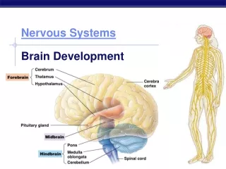
Concept 34.1 Nervous Systems Consist of Neurons and Glia • Networks vary in complexity. • Nerve net—simple network of neurons • Ganglia—neurons organized into clusters, sometimes in pairs, in simple animals • Brain—the largest pair of ganglia, found in animals with complex behavior requiring more information-processing
Figure 34.3 Nervous Systems Vary in Size and Complexity (Part 1)
Figure 34.3 Nervous Systems Vary in Size and Complexity (Part 2)
Figure 34.3 Nervous Systems Vary in Size and Complexity (Part 3)
Concept 34.2 Neurons Generate and Transmit Electrical Signals • Neurons generate changes in membrane potential—the difference in electrical charge across the membrane. • These changes generatenerve impulses, or action potentials. • An action potential is a rapid, large change in membrane potential that travels along an axon and causes release of chemical signals.
Concept 34.2 Neurons Generate and Transmit Electrical Signals • Voltage is a measure of the difference in electrical charge between two points. • Electrical current in solution is carried by ions. Major ions in neurons: • Sodium (Na+) • Potassium (K+) • Calcium (Ca2+) • Chloride (Cl–) • Different concentrations and charges inside and out produce the membrane potential.
Concept 34.2 Neurons Generate and Transmit Electrical Signals • Membrane potentials can be measured in all cells with electrodes. • Resting potential is the membrane potential of a resting, or inactive, neuron. • The resting potential of a membrane is between –60 and –70 millivolts (mV). • The inside of the cell is negative at rest. An action potential allows positive ions to flow in briefly, making the inside of the cell more positive.
Concept 34.2 Neurons Generate and Transmit Electrical Signals • Ion channels and ion transporters in the membrane create the resting and action potentials. • Sodium–potassium pump—moves Na+ ions from inside, exchanges for K+ from outside—establishes concentration gradients • The Na+–K+ pump is an antiporter,or sodium–potassium ATPase, as it requires ATP.
Concept 34.2 Neurons Generate and Transmit Electrical Signals • Potassium channels are open in the resting membrane and are highly permeable to K+ ions—allow leak currents • K+ ions diffuse out of the cell along the concentration gradient and leave behind negative charges within the cell. • K+ ions diffuse back into the cell because of the negative electrical potential. • These two forces acting on K+ are its electrochemical gradient.
Concept 34.2 Neurons Generate and Transmit Electrical Signals • The equilibrium potential is the membrane potential at which the net movement of an ion ceases. • The Nernst equation calculates the value of the equilibrium potential by measuring the concentrations of an ion on both sides of the membrane.
Concept 34.2 Neurons Generate and Transmit Electrical Signals • Some ion channels are “gated”—open and close under certain conditions: • Voltage-gated channels respond to change in voltage across membrane • Chemically-gated channels depend on molecules that bind or alter channel protein • Mechanically-gated channels respond to force applied to membrane
Concept 34.2 Neurons Generate and Transmit Electrical Signals • Gating provides a means for neurons to change their membrane potentials in response to a stimulus. • The membrane is depolarized when Na+ enters the cell and the inside of the neuron becomes less negative. • If gated K+ channels open and K+ leaves, the cell becomes more negative inside and the membrane is hyperpolarized.
Concept 34.2 Neurons Generate and Transmit Electrical Signals • Graded membrane potentials are changes from the resting potential. • Graded potentials are a means of integrating input—the membrane can respond proportionally to depolarization or hyperpolarization.
Concept 34.2 Neurons Generate and Transmit Electrical Signals • Voltage-gated Na+ and K+ channels are responsible for action potentials—sudden, large changes in membrane potential. • At rest most of these channels are closed. • Local depolarization by gated channels in dendrites produces a graded potential. • It spreads to the axon hillock, where Na+ voltage-gated channels are concentrated.
Concept 34.2 Neurons Generate and Transmit Electrical Signals • The membrane in the axon hillock may reach its threshold—5 to 10 mV above resting potential. • Many voltage-gated Na+ channels (activation gates) open quickly and Na+ rushes into the axon. • The influx of positive ions causes more depolarization, the membrane potential is briefly positive, and an action potential occurs.
Concept 34.2 Neurons Generate and Transmit Electrical Signals • The axon quickly returns to resting potential due to two things: • Voltage-gated K+ channels open slowly and stay open longer—K+ moves out • Voltage-gated Na+ channels (inactivation gates) close • Voltage-gated Na+ channels cannot open again during the refractory period—a few milliseconds.
Concept 34.2 Neurons Generate and Transmit Electrical Signals • An action potential is an all-or-none event—positive feedback to voltage-gated Na+ channels ensures the maximum action potential. • An action potential is self-regenerating because it spreads to adjacent membrane regions.
Concept 34.2 Neurons Generate and Transmit Electrical Signals • Axon diameter and myelination by glial cells increase the speed of action potentials in axons. • The nodes of Ranvier are regularly spaced gaps where the axon is not covered by myelin. • Action potentials are generated at the nodes and the positive current flows down the inside of the axon.
Concept 34.2 Neurons Generate and Transmit Electrical Signals • When positive current reaches the next node, the membrane is depolarized—another axon potential is generated. • Action potentials appear to jump from node to node, a form of propagation called saltatory conduction.
Concept 34.3 Neurons Communicate with Other Cells at Synapses • Neurons communicate with other neurons or target cells at synapses. • In a chemical synapse neurotransmitters from a presynaptic cell bind to receptors in a postsynaptic cell. • The synaptic cleft—about 25 nanometers wide—separates the cells.
Concept 34.3 Neurons Communicate with Other Cells at Synapses • In an electrical synapse, cells are joined through gap junctions. • Gap junctions are made of proteins (connexins) that create channels. • Ions flow through the channels—the action potential spreads through the cytoplasm. • These action potentials are fast but do not allow for complex integration of inputs.
Concept 34.3 Neurons Communicate with Other Cells at Synapses • The neuromuscular junction is a chemical synapse between motor neurons and skeletal muscle cells. • An action potential causes voltage-gatedCa+ channels to open in the presynaptic membrane, allowing Ca+ to flow in. • The presynaptic neuron releases acetylcholine(ACh) from its axon terminals (boutons) when vesicles fuse with the membrane.
Concept 34.3 Neurons Communicate with Other Cells at Synapses • The postsynaptic membrane of the muscle cell is the motor end plate. • ACh diffuses across the cleft and binds to ACh receptors on the motor end plate. • These receptors allow Na+ and K+ to flow through, and the increase in Na+ depolarizes the membrane. • If it reaches threshold, more Na+ voltage-gated channels are activated and an action potential is generated.
Concept 34.3 Neurons Communicate with Other Cells at Synapses • The postsynaptic cell must sum the excitatory and inhibitory input. • Summation occurs at the axon hillock, the part of the cell body at the base of the axon. • Spatial summation adds up messages at different synaptic sites. • Temporal summation adds up potentials generated at the same site, over time.